 Movie
Movie Controller


+ Open data

- Basic information
| Entry | Database: PDB / ID: 8wmy | ||||||
|---|---|---|---|---|---|---|---|
| Title | Crystal structure of YqeK in complex with MG and ADP. | ||||||
 Components Components | bis(5'-nucleosyl)-tetraphosphatase (symmetrical) | ||||||
 Keywords Keywords | HYDROLASE / Staphylococcus pyogenes / YqeK / Protein structure / enzymatic activity. | ||||||
| Function / homology |  Function and homology information Function and homology informationbis(5'-nucleosyl)-tetraphosphatase (symmetrical) / hydrolase activity / nucleotide binding / metal ion binding Similarity search - Function | ||||||
| Biological species |  Staphylococcus (bacteria) Staphylococcus (bacteria) | ||||||
| Method |  X-RAY DIFFRACTION / SYNCHROTRON / MOLECULAR REPLACEMENT / Resolution: 1.91 Å X-RAY DIFFRACTION / SYNCHROTRON / MOLECULAR REPLACEMENT / Resolution: 1.91 Å | ||||||
 Authors Authors | Mo, X. | ||||||
| Funding support |  China, 1items China, 1items
| ||||||
 Citation Citation | Journal: To be published Title: Molecular Mechanism of Ap4A Hydrolysis by YqeK of Staphylococcus pyogenes. Authors: Mo, X. | ||||||
| History |
|
- Structure visualization
| Structure viewer | Molecule: MolmilJmol/JSmol |
|---|
- Downloads & links
-Download
| PDBx/mmCIF format | 8wmy.cif.gz | 120.5 KB | Display | PDBx/mmCIF format |
|---|---|---|---|---|
| PDB format | pdb8wmy.ent.gz | 75.5 KB | Display | PDB format |
| PDBx/mmJSON format | 8wmy.json.gz | Tree view | PDBx/mmJSON format | |
| Others |  Other downloads Other downloads |
-Validation report
| Arichive directory | https://data.pdbj.org/pub/pdb/validation_reports/wm/8wmyftp://data.pdbj.org/pub/pdb/validation_reports/wm/8wmy | HTTPS FTP |
|---|
-Related structure data
| Similar structure data |
|---|
-Links
 PDBj
PDBj
- Assembly
| Deposited unit | 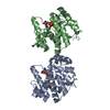
| |||||||||||||||||||||||||||||||||||||||||||||||||||||||||||||
|---|---|---|---|---|---|---|---|---|---|---|---|---|---|---|---|---|---|---|---|---|---|---|---|---|---|---|---|---|---|---|---|---|---|---|---|---|---|---|---|---|---|---|---|---|---|---|---|---|---|---|---|---|---|---|---|---|---|---|---|---|---|---|
| 1 | 
| |||||||||||||||||||||||||||||||||||||||||||||||||||||||||||||
| Unit cell |
| |||||||||||||||||||||||||||||||||||||||||||||||||||||||||||||
| Noncrystallographic symmetry (NCS) | NCS domain:
NCS domain segments: Ens-ID: 1
|
-Components
| #1: Protein | Mass: 22487.834 Da / Num. of mol.: 2 Source method: isolated from a genetically manipulated source Details: Sequence reference for Staphylococcus aureus is not available in UniProt at the time of biocuration. Current sequence reference is from UniProt id A0A4U7IGH6. Source: (gene. exp.) Staphylococcus (bacteria) / Gene: E0F66_02650, SAMEA1711581_00596, SAMEA864267_01112 / Production host: References: UniProt: A0A4U7IGH6, bis(5'-nucleosyl)-tetraphosphatase (symmetrical) #2: Chemical |   Mass: 427.201 Da / Num. of mol.: 2 / Source method: obtained synthetically / Formula: C10H15N5O10P2 / Feature type: SUBJECT OF INVESTIGATION / Comment: ADP, energy-carrying molecule*YM Mass: 427.201 Da / Num. of mol.: 2 / Source method: obtained synthetically / Formula: C10H15N5O10P2 / Feature type: SUBJECT OF INVESTIGATION / Comment: ADP, energy-carrying molecule*YM#3: Chemical | ChemComp-MG /   Mass: 24.305 Da / Num. of mol.: 4 / Source method: obtained synthetically / Formula: Mg / Feature type: SUBJECT OF INVESTIGATION Mass: 24.305 Da / Num. of mol.: 4 / Source method: obtained synthetically / Formula: Mg / Feature type: SUBJECT OF INVESTIGATION#4: Water | ChemComp-HOH / |  Mass: 18.015 Da / Num. of mol.: 311 / Source method: isolated from a natural source / Formula: H2O Mass: 18.015 Da / Num. of mol.: 311 / Source method: isolated from a natural source / Formula: H2OHas ligand of interest | Y | Has protein modification | N | |
|---|
-Experimental details
-Experiment
| Experiment | Method: X-RAY DIFFRACTION / Number of used crystals: 1 |
|---|
- Sample preparation
| Crystal | Density Matthews: 2.55 Å3/Da / Density % sol: 51.82 % |
|---|---|
| Crystal grow | Temperature: 293.15 K / Method: evaporation Details: 12% 2-propannol, 18.5% PEG-4000 and 100 mM sodium HEPES (pH 7.4) |
-Data collection
| Diffraction | Mean temperature: 100 K / Serial crystal experiment: N |
|---|---|
| Diffraction source | Source: SYNCHROTRON / Site: NSRRC  / Beamline: BL13B1 / Wavelength: 1 Å / Beamline: BL13B1 / Wavelength: 1 Å |
| Detector | Type: ADSC QUANTUM 315r / Detector: CCD / Date: Dec 12, 2019 |
| Radiation | Protocol: SINGLE WAVELENGTH / Monochromatic (M) / Laue (L): M / Scattering type: x-ray |
| Radiation wavelength | Wavelength: 1 Å / Relative weight: 1 |
| Reflection | Resolution: 1.91→35.04 Å / Num. obs: 35345 / % possible obs: 98.4 % / Redundancy: 3.7 % / Biso Wilson estimate: 31.17 Å2 / CC1/2: 0.98 / Net I/σ(I): 3 |
| Reflection shell | Resolution: 1.91→1.978 Å / Num. unique obs: 3499 / CC1/2: 0.97 |
- Processing
| Software |
| ||||||||||||||||||||||||||||||||||||||||||||||||||||||||||||||||||||||||||||||||||||||||||||||||||
|---|---|---|---|---|---|---|---|---|---|---|---|---|---|---|---|---|---|---|---|---|---|---|---|---|---|---|---|---|---|---|---|---|---|---|---|---|---|---|---|---|---|---|---|---|---|---|---|---|---|---|---|---|---|---|---|---|---|---|---|---|---|---|---|---|---|---|---|---|---|---|---|---|---|---|---|---|---|---|---|---|---|---|---|---|---|---|---|---|---|---|---|---|---|---|---|---|---|---|---|
| Refinement | Method to determine structure: MOLECULAR REPLACEMENT / Resolution: 1.91→35.04 Å / SU ML: 0.2506 / Cross valid method: FREE R-VALUE / σ(F): 1.37 / Phase error: 26.6335 Stereochemistry target values: GeoStd + Monomer Library + CDL v1.2
| ||||||||||||||||||||||||||||||||||||||||||||||||||||||||||||||||||||||||||||||||||||||||||||||||||
| Solvent computation | Shrinkage radii: 0.9 Å / VDW probe radii: 1.11 Å / Solvent model: FLAT BULK SOLVENT MODEL | ||||||||||||||||||||||||||||||||||||||||||||||||||||||||||||||||||||||||||||||||||||||||||||||||||
| Displacement parameters | Biso mean: 35.8 Å2 | ||||||||||||||||||||||||||||||||||||||||||||||||||||||||||||||||||||||||||||||||||||||||||||||||||
| Refinement step | Cycle: LAST / Resolution: 1.91→35.04 Å
| ||||||||||||||||||||||||||||||||||||||||||||||||||||||||||||||||||||||||||||||||||||||||||||||||||
| Refine LS restraints |
| ||||||||||||||||||||||||||||||||||||||||||||||||||||||||||||||||||||||||||||||||||||||||||||||||||
| LS refinement shell |
|
