+ Open data
- Basic information
| Entry | Database: PDB / ID: 8wmv | |||||||||||||||||||||
|---|---|---|---|---|---|---|---|---|---|---|---|---|---|---|---|---|---|---|---|---|---|---|
| Title | The structure of PSI-14CAC complex at stationary growth phase | |||||||||||||||||||||
 Components Components |
| |||||||||||||||||||||
 Keywords Keywords | PHOTOSYNTHESIS / chlorophyll c / alloxanthin / growth phase | |||||||||||||||||||||
| Function / homology |  Function and homology information Function and homology informationphotosystem I reaction center / photosystem I / photosynthetic electron transport in photosystem I / photosystem I / chlorophyll binding / chloroplast thylakoid membrane / photosynthesis / chloroplast / 4 iron, 4 sulfur cluster binding / electron transfer activity ...photosystem I reaction center / photosystem I / photosynthetic electron transport in photosystem I / photosystem I / chlorophyll binding / chloroplast thylakoid membrane / photosynthesis / chloroplast / 4 iron, 4 sulfur cluster binding / electron transfer activity / oxidoreductase activity / magnesium ion binding / metal ion binding Similarity search - Function | |||||||||||||||||||||
| Biological species |  Rhodomonas salina (eukaryote) Rhodomonas salina (eukaryote) | |||||||||||||||||||||
| Method | ELECTRON MICROSCOPY / single particle reconstruction / cryo EM / Resolution: 2.94 Å | |||||||||||||||||||||
 Authors Authors | Zhang, S.M. / Si, L. / Li, M. | |||||||||||||||||||||
| Funding support |  China, 2items China, 2items
| |||||||||||||||||||||
 Citation Citation |  Journal: Commun Biol / Year: 2024 Journal: Commun Biol / Year: 2024Title: Growth phase-dependent reorganization of cryptophyte photosystem I antennae. Authors: Shumeng Zhang / Long Si / Xiaodong Su / Xuelin Zhao / Xiaomin An / Mei Li /  Abstract: Photosynthetic cryptophytes are eukaryotic algae that utilize membrane-embedded chlorophyll a/c binding proteins (CACs) and lumen-localized phycobiliproteins (PBPs) as their light-harvesting antennae. ...Photosynthetic cryptophytes are eukaryotic algae that utilize membrane-embedded chlorophyll a/c binding proteins (CACs) and lumen-localized phycobiliproteins (PBPs) as their light-harvesting antennae. Cryptophytes go through logarithmic and stationary growth phases, and may adjust their light-harvesting capability according to their particular growth state. How cryptophytes change the type/arrangement of the photosynthetic antenna proteins to regulate their light-harvesting remains unknown. Here we solve four structures of cryptophyte photosystem I (PSI) bound with CACs that show the rearrangement of CACs at different growth phases. We identify a cryptophyte-unique protein, PsaQ, which harbors two chlorophyll molecules. PsaQ specifically binds to the lumenal region of PSI during logarithmic growth phase and may assist the association of PBPs with photosystems and energy transfer from PBPs to photosystems. | |||||||||||||||||||||
| History |
|
- Structure visualization
| Structure viewer | Molecule:  Molmil Molmil Jmol/JSmol Jmol/JSmol |
|---|
- Downloads & links
- Download
| PDBx/mmCIF format |  8wmv.cif.gz 8wmv.cif.gz | 1.3 MB | Display |  PDBx/mmCIF format PDBx/mmCIF format |
|---|---|---|---|---|
| PDB format |  pdb8wmv.ent.gz pdb8wmv.ent.gz | 1.2 MB | Display |  PDB format PDB format |
| PDBx/mmJSON format |  8wmv.json.gz 8wmv.json.gz | Tree view |  PDBx/mmJSON format PDBx/mmJSON format | |
| Others |  Other downloads Other downloads |
-Validation report
| Arichive directory |  https://data.pdbj.org/pub/pdb/validation_reports/wm/8wmv https://data.pdbj.org/pub/pdb/validation_reports/wm/8wmv ftp://data.pdbj.org/pub/pdb/validation_reports/wm/8wmv ftp://data.pdbj.org/pub/pdb/validation_reports/wm/8wmv | HTTPS FTP |
|---|
-Related structure data
| Related structure data |  37659MC  8wm6C  8wmjC  8wmwC  8wnwC M: map data used to model this data C: citing same article ( |
|---|---|
| Similar structure data | Similarity search - Function & homology  F&H Search F&H Search |
- Links
Links
- Assembly
| Deposited unit | 
|
|---|---|
| 1 |
|
- Components
-Photosystem I P700 chlorophyll a apoprotein ... , 2 types, 2 molecules AB
| #1: Protein | Mass: 83431.523 Da / Num. of mol.: 1 / Source method: isolated from a natural source / Source: (natural)  Rhodomonas salina (eukaryote) / References: UniProt: A6MVZ7 Rhodomonas salina (eukaryote) / References: UniProt: A6MVZ7 |
|---|---|
| #2: Protein | Mass: 82069.711 Da / Num. of mol.: 1 / Source method: isolated from a natural source / Source: (natural)  Rhodomonas salina (eukaryote) / References: UniProt: A6MVZ6 Rhodomonas salina (eukaryote) / References: UniProt: A6MVZ6 |
-Protein , 16 types, 18 molecules COscabhmfjelkidgRn
| #3: Protein | Mass: 8759.131 Da / Num. of mol.: 1 / Source method: isolated from a natural source / Source: (natural)  Rhodomonas salina (eukaryote) / References: UniProt: A6MVS8 Rhodomonas salina (eukaryote) / References: UniProt: A6MVS8 | ||||||||||||||||
|---|---|---|---|---|---|---|---|---|---|---|---|---|---|---|---|---|---|
| #11: Protein | Mass: 15133.567 Da / Num. of mol.: 1 / Source method: isolated from a natural source / Source: (natural)  Rhodomonas salina (eukaryote) Rhodomonas salina (eukaryote) | ||||||||||||||||
| #13: Protein | Mass: 27966.930 Da / Num. of mol.: 1 / Source method: isolated from a natural source / Source: (natural)  Rhodomonas salina (eukaryote) Rhodomonas salina (eukaryote) | ||||||||||||||||
| #14: Protein | Mass: 23781.504 Da / Num. of mol.: 1 / Source method: isolated from a natural source / Source: (natural)  Rhodomonas salina (eukaryote) Rhodomonas salina (eukaryote) | ||||||||||||||||
| #15: Protein | Mass: 23330.691 Da / Num. of mol.: 1 / Source method: isolated from a natural source / Source: (natural)  Rhodomonas salina (eukaryote) Rhodomonas salina (eukaryote) | ||||||||||||||||
| #16: Protein | Mass: 23503.363 Da / Num. of mol.: 1 / Source method: isolated from a natural source / Source: (natural)  Rhodomonas salina (eukaryote) Rhodomonas salina (eukaryote) | ||||||||||||||||
| #17: Protein | Mass: 23132.781 Da / Num. of mol.: 1 / Source method: isolated from a natural source / Source: (natural)  Rhodomonas salina (eukaryote) Rhodomonas salina (eukaryote) | ||||||||||||||||
| #18: Protein | Mass: 22272.936 Da / Num. of mol.: 3 / Source method: isolated from a natural source / Source: (natural)  Rhodomonas salina (eukaryote) Rhodomonas salina (eukaryote)#19: Protein | | Mass: 21787.338 Da / Num. of mol.: 1 / Source method: isolated from a natural source / Source: (natural)  Rhodomonas salina (eukaryote) Rhodomonas salina (eukaryote)#20: Protein | | Mass: 25908.721 Da / Num. of mol.: 1 / Source method: isolated from a natural source / Source: (natural)  Rhodomonas salina (eukaryote) Rhodomonas salina (eukaryote)#21: Protein | | Mass: 25588.902 Da / Num. of mol.: 1 / Source method: isolated from a natural source / Source: (natural)  Rhodomonas salina (eukaryote) Rhodomonas salina (eukaryote)#22: Protein | | Mass: 23078.553 Da / Num. of mol.: 1 / Source method: isolated from a natural source / Source: (natural)  Rhodomonas salina (eukaryote) Rhodomonas salina (eukaryote)#23: Protein | | Mass: 22391.033 Da / Num. of mol.: 1 / Source method: isolated from a natural source / Source: (natural)  Rhodomonas salina (eukaryote) Rhodomonas salina (eukaryote)#24: Protein | | Mass: 26979.537 Da / Num. of mol.: 1 / Source method: isolated from a natural source / Source: (natural)  Rhodomonas salina (eukaryote) Rhodomonas salina (eukaryote)#25: Protein | | Mass: 13318.223 Da / Num. of mol.: 1 / Source method: isolated from a natural source / Source: (natural)  Rhodomonas salina (eukaryote) Rhodomonas salina (eukaryote)#26: Protein | | Mass: 22876.436 Da / Num. of mol.: 1 / Source method: isolated from a natural source / Source: (natural)  Rhodomonas salina (eukaryote) Rhodomonas salina (eukaryote) |
-Photosystem I reaction center subunit ... , 8 types, 8 molecules DEFIJLMK
| #4: Protein | Mass: 15585.724 Da / Num. of mol.: 1 / Source method: isolated from a natural source / Source: (natural)  Rhodomonas salina (eukaryote) / References: UniProt: A6MVZ1 Rhodomonas salina (eukaryote) / References: UniProt: A6MVZ1 |
|---|---|
| #5: Protein | Mass: 7309.363 Da / Num. of mol.: 1 / Source method: isolated from a natural source / Source: (natural)  Rhodomonas salina (eukaryote) / References: UniProt: A6MW36 Rhodomonas salina (eukaryote) / References: UniProt: A6MW36 |
| #6: Protein | Mass: 20817.162 Da / Num. of mol.: 1 / Source method: isolated from a natural source / Source: (natural)  Rhodomonas salina (eukaryote) / References: UniProt: A6MVU7 Rhodomonas salina (eukaryote) / References: UniProt: A6MVU7 |
| #7: Protein/peptide | Mass: 3927.708 Da / Num. of mol.: 1 / Source method: isolated from a natural source / Source: (natural)  Rhodomonas salina (eukaryote) / References: UniProt: A6MVV1 Rhodomonas salina (eukaryote) / References: UniProt: A6MVV1 |
| #8: Protein/peptide | Mass: 4974.812 Da / Num. of mol.: 1 / Source method: isolated from a natural source / Source: (natural)  Rhodomonas salina (eukaryote) / References: UniProt: A6MVU6 Rhodomonas salina (eukaryote) / References: UniProt: A6MVU6 |
| #9: Protein | Mass: 16475.051 Da / Num. of mol.: 1 / Source method: isolated from a natural source / Source: (natural)  Rhodomonas salina (eukaryote) / References: UniProt: A6MVV8 Rhodomonas salina (eukaryote) / References: UniProt: A6MVV8 |
| #10: Protein/peptide | Mass: 3318.070 Da / Num. of mol.: 1 / Source method: isolated from a natural source / Source: (natural)  Rhodomonas salina (eukaryote) / References: UniProt: A6MVT4 Rhodomonas salina (eukaryote) / References: UniProt: A6MVT4 |
| #12: Protein | Mass: 8715.256 Da / Num. of mol.: 1 / Source method: isolated from a natural source / Source: (natural)  Rhodomonas salina (eukaryote) / References: UniProt: A6MVS9 Rhodomonas salina (eukaryote) / References: UniProt: A6MVS9 |
-Sugars , 3 types, 6 molecules 
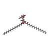
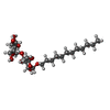


| #31: Sugar | ChemComp-LMT / #33: Sugar | ChemComp-DGD / | #38: Sugar | ChemComp-LMU / | |
|---|
-Non-polymers , 10 types, 538 molecules 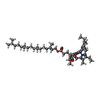
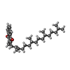


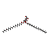

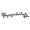


| #27: Chemical | ChemComp-CLA / #28: Chemical | #29: Chemical | ChemComp-LHG / #30: Chemical | ChemComp-WVN / Mass: 536.873 Da / Num. of mol.: 29 / Source method: obtained synthetically / Formula: C40H56 / Feature type: SUBJECT OF INVESTIGATION #32: Chemical | #34: Chemical | ChemComp-LMG / #35: Chemical | ChemComp-II0 / ( #36: Chemical | ChemComp-IHT / ( #37: Chemical | ChemComp-KC2 / #39: Water | ChemComp-HOH / | |
|---|
-Details
| Has ligand of interest | Y |
|---|---|
| Has protein modification | Y |
-Experimental details
-Experiment
| Experiment | Method: ELECTRON MICROSCOPY |
|---|---|
| EM experiment | Aggregation state: PARTICLE / 3D reconstruction method: single particle reconstruction |
- Sample preparation
| Component | Name: PSI-14CAC(L-phase) / Type: COMPLEX / Entity ID: #1-#26 / Source: NATURAL |
|---|---|
| Source (natural) | Organism:  Rhodomonas salina (eukaryote) Rhodomonas salina (eukaryote) |
| Buffer solution | pH: 7.5 |
| Specimen | Embedding applied: NO / Shadowing applied: NO / Staining applied: NO / Vitrification applied: YES |
| Vitrification | Cryogen name: ETHANE |
- Electron microscopy imaging
| Experimental equipment |  Model: Titan Krios / Image courtesy: FEI Company |
|---|---|
| Microscopy | Model: FEI TITAN KRIOS |
| Electron gun | Electron source:  FIELD EMISSION GUN / Accelerating voltage: 300 kV / Illumination mode: FLOOD BEAM FIELD EMISSION GUN / Accelerating voltage: 300 kV / Illumination mode: FLOOD BEAM |
| Electron lens | Mode: BRIGHT FIELD / Nominal defocus max: 2200 nm / Nominal defocus min: 1200 nm |
| Image recording | Electron dose: 60 e/Å2 / Film or detector model: GATAN K2 QUANTUM (4k x 4k) |
- Processing
| CTF correction | Type: PHASE FLIPPING AND AMPLITUDE CORRECTION | ||||||||||||||||||||||||
|---|---|---|---|---|---|---|---|---|---|---|---|---|---|---|---|---|---|---|---|---|---|---|---|---|---|
| 3D reconstruction | Resolution: 2.94 Å / Resolution method: FSC 0.143 CUT-OFF / Num. of particles: 42423 / Symmetry type: POINT | ||||||||||||||||||||||||
| Refinement | Cross valid method: NONE Stereochemistry target values: GeoStd + Monomer Library + CDL v1.2 | ||||||||||||||||||||||||
| Displacement parameters | Biso mean: 33.22 Å2 | ||||||||||||||||||||||||
| Refine LS restraints |
|
 Movie
Movie Controller


 PDBj
PDBj


