National Natural Science Foundation of China (NSFC)
China
Citation
Journal: Nat Commun / Year: 2025 Title: Cryo-EM structure of the β-1,3-glucan synthase FKS1-Rho1 complex. Authors: Jialu Li / Huayi Liu / Jian Li / Juxiu Liu / Xinli Dai / Angqi Zhu / Qingjie Xiao / Wenyu Qian / Honghao Li / Li Guo / Chuangye Yan / Dong Deng / Yunzi Luo / Xiang Wang / Abstract: β-1,3 Glucan synthase (GS) is essential for fungal cell wall biosynthesis. The GS holoenzyme comprises the glycosyltransferase FKS1 and its regulatory factor Rho1, a small GTPase. However, the ...β-1,3 Glucan synthase (GS) is essential for fungal cell wall biosynthesis. The GS holoenzyme comprises the glycosyltransferase FKS1 and its regulatory factor Rho1, a small GTPase. However, the mechanism by which Rho1 activates FKS1 in a GTP-dependent manner remains unclear. Here, we present two cryo-EM structures of FKS1, apo and in complex with Rho1. FKS1 adopts a cellulose synthase-like conformation. The interaction between Rho1 and FKS1 is enhanced in the presence of GTPγS. Rho1 is positioned within a pocket between the glycosyltransferase domain of FKS1 (GT domain) and the transmembrane helix spanning TM7-15. Comparison of the two structures reveals extensive conformational changes within FKS1. These alterations suggest that Rho1's GTP/GDP cycling may act as a molecular pump, promoting a dynamic transition between the resting and active states of FKS1. Notably, Rho1 triggers FKS1 conformational changes that may push the growing glucan chain into FKS1's transmembrane channel, thereby facilitating β-1,3-glucan elongation.
1,3-beta-glucansynthasecomponentFKS1 / 1 / 3-beta-D-glucan-UDP glucosyltransferase / Calcineurin dependent protein 1 / Calcofluor white ...1 / 3-beta-D-glucan-UDP glucosyltransferase / Calcineurin dependent protein 1 / Calcofluor white hypersensitivity protein 53 / Echinocandin target gene protein 1 / FK506 sensitivity protein 1 / Glucan synthase of cerevisiae protein 1 / Papulacandin B resistance protein 1
Mass: 215076.156 Da / Num. of mol.: 1 / Source method: isolated from a natural source / Source: (natural) Saccharomyces cerevisiae (brewer's yeast) / References: UniProt: P38631, 1,3-beta-glucan synthase
Has protein modification
Y
-
Experimental details
-
Experiment
Experiment
Method: ELECTRON MICROSCOPY
EM experiment
Aggregation state: PARTICLE / 3D reconstruction method: single particle reconstruction
-
Sample preparation
Component
Name: 1,3-beta-glucan synthase component FKS1 in apo state / Type: COMPLEX / Entity ID: all / Source: NATURAL
In the structure databanks used in Yorodumi, some data are registered as the other names, "COVID-19 virus" and "2019-nCoV". Here are the details of the virus and the list of structure data.
Jan 31, 2019. EMDB accession codes are about to change! (news from PDBe EMDB page)
EMDB accession codes are about to change! (news from PDBe EMDB page)
The allocation of 4 digits for EMDB accession codes will soon come to an end. Whilst these codes will remain in use, new EMDB accession codes will include an additional digit and will expand incrementally as the available range of codes is exhausted. The current 4-digit format prefixed with “EMD-” (i.e. EMD-XXXX) will advance to a 5-digit format (i.e. EMD-XXXXX), and so on. It is currently estimated that the 4-digit codes will be depleted around Spring 2019, at which point the 5-digit format will come into force.
The EM Navigator/Yorodumi systems omit the EMD- prefix.
Related info.:Q: What is EMD? / ID/Accession-code notation in Yorodumi/EM Navigator
Yorodumi is a browser for structure data from EMDB, PDB, SASBDB, etc.
This page is also the successor to EM Navigator detail page, and also detail information page/front-end page for Omokage search.
The word "yorodu" (or yorozu) is an old Japanese word meaning "ten thousand". "mi" (miru) is to see.
Related info.:EMDB / PDB / SASBDB / Comparison of 3 databanks / Yorodumi Search / Aug 31, 2016. New EM Navigator & Yorodumi / Yorodumi Papers / Jmol/JSmol / Function and homology information / Changes in new EM Navigator and Yorodumi
 Movie
Movie Controller


 Open data
 Basic information
 Components
 Keywords
 Function and homology information
 Authors
 China, 1items
  Citation
 Structure visualization
 Downloads & links
 Other downloads
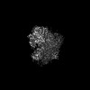


 PDBj
PDBj Assembly
 Sample preparation
 Electron microscopy imaging
 FIELD EMISSION GUN / Accelerating voltage: 300 kV / Illumination mode: FLOOD BEAM
 Processing