 Movie
Movie Controller

[English] 日本語
 Yorodumi
- PDB-8wl1: The crystal structure of D-mandelate dehydrogenase from Lactobaci... -
+ Open data

- Basic information
| Entry | Database: PDB / ID: 8wl1 | ||||||
|---|---|---|---|---|---|---|---|
| Title | The crystal structure of D-mandelate dehydrogenase from Lactobacillus brevis | ||||||
 Components Components | 2-dehydropantoate 2-reductase | ||||||
 Keywords Keywords | OXIDOREDUCTASE / D-Mandelate dehydrogenase 2-Ketopantoate reductase Crystal structure | ||||||
| Function / homology |  Function and homology information Function and homology information2-dehydropantoate 2-reductase / 2-dehydropantoate 2-reductase activity / pantothenate biosynthetic process / NADP binding / cytoplasm Similarity search - Function | ||||||
| Biological species |  Levilactobacillus brevis (bacteria) Levilactobacillus brevis (bacteria) | ||||||
| Method |  X-RAY DIFFRACTION / SYNCHROTRON / MOLECULAR REPLACEMENT / Resolution: 1.75 Å X-RAY DIFFRACTION / SYNCHROTRON / MOLECULAR REPLACEMENT / Resolution: 1.75 Å | ||||||
 Authors Authors | Liu, F. / Rao, Z.M. | ||||||
| Funding support |  China, 1items China, 1items
| ||||||
 Citation Citation | Journal: To Be Published Title: The crystal structure of D-mandelate dehydrogenase from Lactobacillus brevis Authors: Liu, F. / Rao, Z.M. | ||||||
| History |
|
- Structure visualization
| Structure viewer | Molecule: MolmilJmol/JSmol |
|---|
- Downloads & links
-Download
| PDBx/mmCIF format | 8wl1.cif.gz | 74.7 KB | Display | PDBx/mmCIF format |
|---|---|---|---|---|
| PDB format | pdb8wl1.ent.gz | 54.3 KB | Display | PDB format |
| PDBx/mmJSON format | 8wl1.json.gz | Tree view | PDBx/mmJSON format | |
| Others |  Other downloads Other downloads |
-Validation report
| Summary document | 8wl1_validation.pdf.gz | 420.6 KB | Display | wwPDB validaton report |
|---|---|---|---|---|
| Full document | 8wl1_full_validation.pdf.gz | 424.6 KB | Display | |
| Data in XML | 8wl1_validation.xml.gz | 15.8 KB | Display | |
| Data in CIF | 8wl1_validation.cif.gz | 21.3 KB | Display | |
| Arichive directory | https://data.pdbj.org/pub/pdb/validation_reports/wl/8wl1ftp://data.pdbj.org/pub/pdb/validation_reports/wl/8wl1 | HTTPS FTP |
-Related structure data
| Similar structure data |
|---|
-Links
 PDBj
PDBj


- Assembly
| Deposited unit | 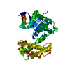
| ||||||||
|---|---|---|---|---|---|---|---|---|---|
| 1 | 
| ||||||||
| Unit cell |
| ||||||||
| Components on special symmetry positions |
|
-Components
| #1: Protein | Mass: 33342.637 Da / Num. of mol.: 1 Source method: isolated from a genetically manipulated source Source: (gene. exp.) Levilactobacillus brevis (bacteria) / Gene: CNR30_12720, UCCLB556_2064, LbMDH / Production host: References: UniProt: A0A7Z6MKR0, 2-dehydropantoate 2-reductase |
|---|---|
| #2: Water | ChemComp-HOH /  Mass: 18.015 Da / Num. of mol.: 136 / Source method: isolated from a natural source / Formula: H2O Mass: 18.015 Da / Num. of mol.: 136 / Source method: isolated from a natural source / Formula: H2O |
-Experimental details
-Experiment
| Experiment | Method: X-RAY DIFFRACTION / Number of used crystals: 1 |
|---|
- Sample preparation
| Crystal | Density Matthews: 2.4 Å3/Da / Density % sol: 48.82 % |
|---|---|
| Crystal grow | Temperature: 289.15 K / Method: vapor diffusion, hanging drop Details: 0.3 M NaCl, 0.05M Tris pH 7.5, 30% Polyethylene glycol 335 |
-Data collection
| Diffraction | Mean temperature: 100 K / Serial crystal experiment: N |
|---|---|
| Diffraction source | Source: SYNCHROTRON / Site: SSRF / Beamline: BL19U1 / Wavelength: 0.9786 Å |
| Detector | Type: DECTRIS PILATUS3 6M / Detector: PIXEL / Date: Nov 22, 2020 |
| Radiation | Protocol: SINGLE WAVELENGTH / Monochromatic (M) / Laue (L): M / Scattering type: x-ray |
| Radiation wavelength | Wavelength: 0.9786 Å / Relative weight: 1 |
| Reflection | Resolution: 1.75→34.92 Å / Num. obs: 32473 / % possible obs: 99.1 % / Redundancy: 13.1 % / Biso Wilson estimate: 29.01 Å2 / CC1/2: 1 / Net I/σ(I): 29.5 |
| Reflection shell | Resolution: 1.75→1.78 Å / Num. unique obs: 32473 / CC1/2: 1 |
- Processing
| Software |
| ||||||||||||||||||||||||||||||||||||||||||||||||||||||||||||||||||||||||||||||||||||||||||||||||||||||||||||||||||||||||||||||||||||||||||||||||||||||||||||||||||||||||||||||||||||||
|---|---|---|---|---|---|---|---|---|---|---|---|---|---|---|---|---|---|---|---|---|---|---|---|---|---|---|---|---|---|---|---|---|---|---|---|---|---|---|---|---|---|---|---|---|---|---|---|---|---|---|---|---|---|---|---|---|---|---|---|---|---|---|---|---|---|---|---|---|---|---|---|---|---|---|---|---|---|---|---|---|---|---|---|---|---|---|---|---|---|---|---|---|---|---|---|---|---|---|---|---|---|---|---|---|---|---|---|---|---|---|---|---|---|---|---|---|---|---|---|---|---|---|---|---|---|---|---|---|---|---|---|---|---|---|---|---|---|---|---|---|---|---|---|---|---|---|---|---|---|---|---|---|---|---|---|---|---|---|---|---|---|---|---|---|---|---|---|---|---|---|---|---|---|---|---|---|---|---|---|---|---|---|---|
| Refinement | Method to determine structure: MOLECULAR REPLACEMENT / Resolution: 1.75→34.92 Å / Cor.coef. Fo:Fc: 0.951 / Cor.coef. Fo:Fc free: 0.927 / SU B: 2.689 / SU ML: 0.087 / Cross valid method: THROUGHOUT / ESU R: 0.133 / ESU R Free: 0.129 / Stereochemistry target values: MAXIMUM LIKELIHOOD / Details: HYDROGENS HAVE BEEN ADDED IN THE RIDING POSITIONS
| ||||||||||||||||||||||||||||||||||||||||||||||||||||||||||||||||||||||||||||||||||||||||||||||||||||||||||||||||||||||||||||||||||||||||||||||||||||||||||||||||||||||||||||||||||||||
| Solvent computation | Ion probe radii: 0.8 Å / Shrinkage radii: 0.8 Å / VDW probe radii: 1.2 Å / Solvent model: MASK | ||||||||||||||||||||||||||||||||||||||||||||||||||||||||||||||||||||||||||||||||||||||||||||||||||||||||||||||||||||||||||||||||||||||||||||||||||||||||||||||||||||||||||||||||||||||
| Displacement parameters | Biso mean: 33.067 Å2
| ||||||||||||||||||||||||||||||||||||||||||||||||||||||||||||||||||||||||||||||||||||||||||||||||||||||||||||||||||||||||||||||||||||||||||||||||||||||||||||||||||||||||||||||||||||||
| Refinement step | Cycle: 1 / Resolution: 1.75→34.92 Å
| ||||||||||||||||||||||||||||||||||||||||||||||||||||||||||||||||||||||||||||||||||||||||||||||||||||||||||||||||||||||||||||||||||||||||||||||||||||||||||||||||||||||||||||||||||||||
| Refine LS restraints |
|