 Movie
Movie Controller


+ Open data

- Basic information
| Entry | Database: PDB / ID: 8wku | ||||||
|---|---|---|---|---|---|---|---|
| Title | Complex structure of MjHKU4r-CoV-1 spike RBD bound to human CD26 | ||||||
 Components Components |
| ||||||
 Keywords Keywords | VIRAL PROTEIN/HYDROLASE / MjHKU4r-CoV-1 / spike RBD / human CD26 / VIRAL PROTEIN-HYDROLASE complex | ||||||
| Function / homology |  Function and homology information Function and homology informationglucagon processing / negative regulation of neutrophil chemotaxis / regulation of cell-cell adhesion mediated by integrin / Synthesis, secretion, and inactivation of Glucose-dependent Insulinotropic Polypeptide (GIP) / dipeptidyl-peptidase IV / negative regulation of extracellular matrix disassembly / chemorepellent activity / intercellular canaliculus / psychomotor behavior / dipeptidyl-peptidase activity ...glucagon processing / negative regulation of neutrophil chemotaxis / regulation of cell-cell adhesion mediated by integrin / Synthesis, secretion, and inactivation of Glucose-dependent Insulinotropic Polypeptide (GIP) / dipeptidyl-peptidase IV / negative regulation of extracellular matrix disassembly / chemorepellent activity / intercellular canaliculus / psychomotor behavior / dipeptidyl-peptidase activity / peptide hormone processing / lamellipodium membrane / locomotory exploration behavior / aminopeptidase activity / endocytic vesicle / endothelial cell migration / behavioral fear response / T cell costimulation / receptor-mediated endocytosis of virus by host cell / serine-type peptidase activity / T cell activation / Synthesis, secretion, and inactivation of Glucagon-like Peptide-1 (GLP-1) / lamellipodium / virus receptor activity / protease binding / membrane fusion / response to hypoxia / receptor-mediated virion attachment to host cell / cell adhesion / apical plasma membrane / membrane raft / signaling receptor binding / serine-type endopeptidase activity / lysosomal membrane / focal adhesion / positive regulation of cell population proliferation / symbiont entry into host cell / cell surface / protein homodimerization activity / proteolysis / extracellular exosome / extracellular region / membrane / identical protein binding / plasma membrane Similarity search - Function | ||||||
| Biological species |  Homo sapiens (human) Homo sapiens (human) | ||||||
| Method |  X-RAY DIFFRACTION / SYNCHROTRON / MOLECULAR REPLACEMENT / Resolution: 3.5 Å X-RAY DIFFRACTION / SYNCHROTRON / MOLECULAR REPLACEMENT / Resolution: 3.5 Å | ||||||
 Authors Authors | Zhao, Z.N. / Chai, Y. / Gao, F. | ||||||
| Funding support |  China, 1items China, 1items
| ||||||
 Citation Citation | Journal: Embo Rep. / Year: 2024 Title: Molecular basis for receptor recognition and broad host tropism for merbecovirus MjHKU4r-CoV-1. Authors: Zhao, Z. / Li, X. / Chai, Y. / Liu, Z. / Wang, Q. / Gao, G.F. | ||||||
| History |
|
- Structure visualization
| Structure viewer | Molecule: MolmilJmol/JSmol |
|---|
- Downloads & links
-Download
| PDBx/mmCIF format | 8wku.cif.gz | 234.5 KB | Display | PDBx/mmCIF format |
|---|---|---|---|---|
| PDB format | pdb8wku.ent.gz | 169.2 KB | Display | PDB format |
| PDBx/mmJSON format | 8wku.json.gz | Tree view | PDBx/mmJSON format | |
| Others |  Other downloads Other downloads |
-Validation report
| Arichive directory | https://data.pdbj.org/pub/pdb/validation_reports/wk/8wkuftp://data.pdbj.org/pub/pdb/validation_reports/wk/8wku | HTTPS FTP |
|---|
-Related structure data
| Related structure data | 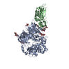 4qzvS S: Starting model for refinement |
|---|---|
| Similar structure data |
-Links
 PDBj
PDBj


- Assembly
| Deposited unit | 
| ||||||||||||
|---|---|---|---|---|---|---|---|---|---|---|---|---|---|
| 1 | 
| ||||||||||||
| Unit cell |
|
-Components
-Protein , 2 types, 2 molecules AB
| #1: Protein | Mass: 85291.508 Da / Num. of mol.: 1 Source method: isolated from a genetically manipulated source Source: (gene. exp.) Homo sapiens (human) / Gene: DPP4, ADCP2, CD26 / Production host: Homo sapiens (human) / References: UniProt: P27487 |
|---|---|
| #2: Protein | Mass: 27251.791 Da / Num. of mol.: 1 Source method: isolated from a genetically manipulated source Details: Manis javanica HKU4-related coronavirus / Source: (gene. exp.)   Spodoptera frugiperda (fall armyworm) Spodoptera frugiperda (fall armyworm) |
-Sugars , 7 types, 10 molecules 
| #3: Polysaccharide | beta-D-mannopyranose-(1-4)-2-acetamido-2-deoxy-beta-D-glucopyranose-(1-4)-[alpha-L-fucopyranose-(1- ...beta-D-mannopyranose-(1-4)-2-acetamido-2-deoxy-beta-D-glucopyranose-(1-4)-[alpha-L-fucopyranose-(1-6)]2-acetamido-2-deoxy-beta-D-glucopyranose Source method: isolated from a genetically manipulated source | ||||||||
|---|---|---|---|---|---|---|---|---|---|
| #4: Polysaccharide | alpha-D-mannopyranose-(1-3)-[alpha-D-mannopyranose-(1-6)]beta-D-mannopyranose-(1-4)-2-acetamido-2- ...alpha-D-mannopyranose-(1-3)-[alpha-D-mannopyranose-(1-6)]beta-D-mannopyranose-(1-4)-2-acetamido-2-deoxy-beta-D-glucopyranose-(1-4)-[alpha-L-fucopyranose-(1-6)]2-acetamido-2-deoxy-beta-D-glucopyranose Source method: isolated from a genetically manipulated source | ||||||||
| #5: Polysaccharide | Source method: isolated from a genetically manipulated source #6: Polysaccharide | alpha-D-mannopyranose-(1-2)-alpha-D-mannopyranose-(1-2)-alpha-D-mannopyranose-(1-3)-[alpha-D- ...alpha-D-mannopyranose-(1-2)-alpha-D-mannopyranose-(1-2)-alpha-D-mannopyranose-(1-3)-[alpha-D-mannopyranose-(1-6)-alpha-D-mannopyranose-(1-6)]beta-D-mannopyranose-(1-4)-2-acetamido-2-deoxy-beta-D-glucopyranose-(1-4)-2-acetamido-2-deoxy-beta-D-glucopyranose | Source method: isolated from a genetically manipulated source #7: Polysaccharide | alpha-D-mannopyranose-(1-6)-beta-D-mannopyranose-(1-4)-2-acetamido-2-deoxy-beta-D-glucopyranose-(1- ...alpha-D-mannopyranose-(1-6)-beta-D-mannopyranose-(1-4)-2-acetamido-2-deoxy-beta-D-glucopyranose-(1-4)-[alpha-L-fucopyranose-(1-6)]2-acetamido-2-deoxy-beta-D-glucopyranose | Source method: isolated from a genetically manipulated source #8: Polysaccharide | beta-D-mannopyranose-(1-4)-2-acetamido-2-deoxy-beta-D-glucopyranose-(1-4)-2-acetamido-2-deoxy-beta- ...beta-D-mannopyranose-(1-4)-2-acetamido-2-deoxy-beta-D-glucopyranose-(1-4)-2-acetamido-2-deoxy-beta-D-glucopyranose | Source method: isolated from a genetically manipulated source #9: Sugar |  Type: D-saccharide, beta linking / Mass: 221.208 Da / Num. of mol.: 3 / Source method: obtained synthetically / Formula: C8H15NO6 / Feature type: SUBJECT OF INVESTIGATION Type: D-saccharide, beta linking / Mass: 221.208 Da / Num. of mol.: 3 / Source method: obtained synthetically / Formula: C8H15NO6 / Feature type: SUBJECT OF INVESTIGATION |
-Details
| Has ligand of interest | Y |
|---|---|
| Has protein modification | Y |
-Experimental details
-Experiment
| Experiment | Method: X-RAY DIFFRACTION / Number of used crystals: 1 |
|---|
- Sample preparation
| Crystal | Density Matthews: 5.12 Å3/Da / Density % sol: 75.98 % |
|---|---|
| Crystal grow | Temperature: 291 K / Method: vapor diffusion, sitting drop / pH: 8.5 Details: 0.1 M Amino acids, 0.1 M buffer system 3 (Tris base; Bicine) and 30% v/v EDO_P8K |
-Data collection
| Diffraction | Mean temperature: 100 K / Serial crystal experiment: N |
|---|---|
| Diffraction source | Source: SYNCHROTRON / Site: SSRF / Beamline: BL02U1 / Wavelength: 0.9792 Å |
| Detector | Type: DECTRIS EIGER X 9M / Detector: PIXEL / Date: May 22, 2023 |
| Radiation | Protocol: SINGLE WAVELENGTH / Monochromatic (M) / Laue (L): M / Scattering type: x-ray |
| Radiation wavelength | Wavelength: 0.9792 Å / Relative weight: 1 |
| Reflection | Resolution: 3.5→50 Å / Num. obs: 54034 / % possible obs: 95.9 % / Redundancy: 7 % / CC1/2: 0.995 / Net I/σ(I): 10.31 |
| Reflection shell | Resolution: 3.5→3.71 Å / Num. unique obs: 6746 / CC1/2: 0.625 |
- Processing
| Software |
| |||||||||||||||||||||||||||||||||||||||||||||||||||||||||||||||||||||||||||||
|---|---|---|---|---|---|---|---|---|---|---|---|---|---|---|---|---|---|---|---|---|---|---|---|---|---|---|---|---|---|---|---|---|---|---|---|---|---|---|---|---|---|---|---|---|---|---|---|---|---|---|---|---|---|---|---|---|---|---|---|---|---|---|---|---|---|---|---|---|---|---|---|---|---|---|---|---|---|---|
| Refinement | Method to determine structure: MOLECULAR REPLACEMENT Starting model: 4QZV Resolution: 3.5→45.78 Å / Cross valid method: FREE R-VALUE / σ(F): 268.92 / Phase error: 25.1102 Stereochemistry target values: GeoStd + Monomer Library + CDL v1.2
| |||||||||||||||||||||||||||||||||||||||||||||||||||||||||||||||||||||||||||||
| Solvent computation | Shrinkage radii: 0.9 Å / VDW probe radii: 1.1 Å / Solvent model: FLAT BULK SOLVENT MODEL | |||||||||||||||||||||||||||||||||||||||||||||||||||||||||||||||||||||||||||||
| Displacement parameters | Biso mean: 107.39 Å2 | |||||||||||||||||||||||||||||||||||||||||||||||||||||||||||||||||||||||||||||
| Refinement step | Cycle: LAST / Resolution: 3.5→45.78 Å
| |||||||||||||||||||||||||||||||||||||||||||||||||||||||||||||||||||||||||||||
| Refine LS restraints |
| |||||||||||||||||||||||||||||||||||||||||||||||||||||||||||||||||||||||||||||
| LS refinement shell |
|
