 Movie
Movie Controller

[English] 日本語
 Yorodumi
- PDB-8wk1: Bovine trypsin in complex with Durio zibethinus trypsin inhibitor... -
+ Open data

- Basic information
| Entry | Database: PDB / ID: 8wk1 | ||||||||||||
|---|---|---|---|---|---|---|---|---|---|---|---|---|---|
| Title | Bovine trypsin in complex with Durio zibethinus trypsin inhibitor DzTI-4 | ||||||||||||
 Components Components |
| ||||||||||||
 Keywords Keywords | PLANT PROTEIN / Kunitz-type trypsin inhibitor / seed protein / Durio zibethinus / bovine trypsin | ||||||||||||
| Function / homology |  Function and homology information Function and homology informationendopeptidase inhibitor activity / trypsin / serpin family protein binding / serine protease inhibitor complex / digestion / endopeptidase activity / serine-type endopeptidase activity / proteolysis / extracellular space / metal ion binding Similarity search - Function | ||||||||||||
| Biological species |  Durio zibethinus (durian) Durio zibethinus (durian) | ||||||||||||
| Method |  X-RAY DIFFRACTION / SYNCHROTRON / MOLECULAR REPLACEMENT / Resolution: 2 Å X-RAY DIFFRACTION / SYNCHROTRON / MOLECULAR REPLACEMENT / Resolution: 2 Å | ||||||||||||
 Authors Authors | Deentanya, P. / Wangkanont, K. | ||||||||||||
| Funding support |  Thailand, 3items Thailand, 3items
| ||||||||||||
 Citation Citation | Journal: Protein Sci. / Year: 2024 Title: Kunitz-type trypsin inhibitor from durian (Durio zibethinus) employs a distinct loop for trypsin inhibition. Authors: Deetanya, P. / Limsardsanakij, K. / Sabat, G. / Pattaradilokrat, S. / Chaisuekul, C. / Wangkanont, K. | ||||||||||||
| History |
|
- Structure visualization
| Structure viewer | Molecule: MolmilJmol/JSmol |
|---|
- Downloads & links
-Download
| PDBx/mmCIF format | 8wk1.cif.gz | 196.5 KB | Display | PDBx/mmCIF format |
|---|---|---|---|---|
| PDB format | pdb8wk1.ent.gz | 139.3 KB | Display | PDB format |
| PDBx/mmJSON format | 8wk1.json.gz | Tree view | PDBx/mmJSON format | |
| Others |  Other downloads Other downloads |
-Validation report
| Summary document | 8wk1_validation.pdf.gz | 486.3 KB | Display | wwPDB validaton report |
|---|---|---|---|---|
| Full document | 8wk1_full_validation.pdf.gz | 492.1 KB | Display | |
| Data in XML | 8wk1_validation.xml.gz | 43.5 KB | Display | |
| Data in CIF | 8wk1_validation.cif.gz | 58.3 KB | Display | |
| Arichive directory | https://data.pdbj.org/pub/pdb/validation_reports/wk/8wk1ftp://data.pdbj.org/pub/pdb/validation_reports/wk/8wk1 | HTTPS FTP |
-Related structure data
| Related structure data |  8we5C  8wfoC  8whcC  8wi1C  8winC 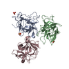 8wioC  8wkbC  8wq6C C: citing same article ( |
|---|---|
| Similar structure data |
-Links
 PDBj
PDBj

- Assembly
| Deposited unit | 
| ||||||||||||
|---|---|---|---|---|---|---|---|---|---|---|---|---|---|
| 1 |
| ||||||||||||
| Unit cell |
|
-Components
-Protein , 2 types, 4 molecules ACBD
| #1: Protein | Mass: 23324.287 Da / Num. of mol.: 2 / Source method: isolated from a natural source / Source: (natural) #2: Protein | Mass: 21673.354 Da / Num. of mol.: 2 Source method: isolated from a genetically manipulated source Source: (gene. exp.) Durio zibethinus (durian) / Strain: Chanee / Gene: LOC111287540 / Plasmid: pET32b / Production host:  |
|---|
-Non-polymers , 4 types, 591 molecules 


| #3: Chemical | ChemComp-SO4 /  Mass: 96.063 Da / Num. of mol.: 4 / Source method: obtained synthetically / Formula: SO4 Mass: 96.063 Da / Num. of mol.: 4 / Source method: obtained synthetically / Formula: SO4#4: Chemical | ChemComp-GOL /  Mass: 92.094 Da / Num. of mol.: 11 / Source method: obtained synthetically / Formula: C3H8O3 Mass: 92.094 Da / Num. of mol.: 11 / Source method: obtained synthetically / Formula: C3H8O3#5: Chemical |  Mass: 59.044 Da / Num. of mol.: 3 / Source method: obtained synthetically / Formula: C2H3O2 Mass: 59.044 Da / Num. of mol.: 3 / Source method: obtained synthetically / Formula: C2H3O2#6: Water | ChemComp-HOH / | Mass: 18.015 Da / Num. of mol.: 573 / Source method: isolated from a natural source / Formula: H2O |
|---|
-Details
| Has ligand of interest | N |
|---|---|
| Has protein modification | Y |
-Experimental details
-Experiment
| Experiment | Method: X-RAY DIFFRACTION / Number of used crystals: 1 |
|---|
- Sample preparation
| Crystal | Density Matthews: 2.08 Å3/Da / Density % sol: 40.88 % |
|---|---|
| Crystal grow | Temperature: 289 K / Method: vapor diffusion, hanging drop / pH: 4.5 Details: 100 mM sodium acetate, 200 mM ammonium sulfate, 25% PEG 4,000 |
-Data collection
| Diffraction | Mean temperature: 100 K / Serial crystal experiment: N |
|---|---|
| Diffraction source | Source: SYNCHROTRON / Site: APS  / Beamline: 21-ID-F / Wavelength: 0.97872 Å / Beamline: 21-ID-F / Wavelength: 0.97872 Å |
| Detector | Type: RAYONIX MX-300 / Detector: CCD / Date: Sep 17, 2022 |
| Radiation | Monochromator: C(111) / Protocol: SINGLE WAVELENGTH / Monochromatic (M) / Laue (L): M / Scattering type: x-ray |
| Radiation wavelength | Wavelength: 0.97872 Å / Relative weight: 1 |
| Reflection | Resolution: 2→29.76 Å / Num. obs: 51078 / % possible obs: 99.2 % / Redundancy: 7.4 % / Biso Wilson estimate: 28.12 Å2 / CC1/2: 0.997 / Rmerge(I) obs: 0.115 / Rpim(I) all: 0.045 / Rrim(I) all: 0.124 / Χ2: 0.98 / Net I/σ(I): 11.2 |
| Reflection shell | Resolution: 2→2.05 Å / Redundancy: 7.6 % / Rmerge(I) obs: 0.922 / Mean I/σ(I) obs: 2.2 / Num. unique obs: 3718 / CC1/2: 0.788 / Rpim(I) all: 0.356 / Rrim(I) all: 0.989 / Χ2: 1 / % possible all: 98.6 |
- Processing
| Software |
| |||||||||||||||||||||||||||||||||||||||||||||||||||||||||||||||||||||||||||||||||||||||||||||||||||||||||||||||||||||||||||||||||||||
|---|---|---|---|---|---|---|---|---|---|---|---|---|---|---|---|---|---|---|---|---|---|---|---|---|---|---|---|---|---|---|---|---|---|---|---|---|---|---|---|---|---|---|---|---|---|---|---|---|---|---|---|---|---|---|---|---|---|---|---|---|---|---|---|---|---|---|---|---|---|---|---|---|---|---|---|---|---|---|---|---|---|---|---|---|---|---|---|---|---|---|---|---|---|---|---|---|---|---|---|---|---|---|---|---|---|---|---|---|---|---|---|---|---|---|---|---|---|---|---|---|---|---|---|---|---|---|---|---|---|---|---|---|---|---|
| Refinement | Method to determine structure: MOLECULAR REPLACEMENT / Resolution: 2→29.76 Å / SU ML: 0.2027 / Cross valid method: FREE R-VALUE / σ(F): 1.33 / Phase error: 22.8928 Stereochemistry target values: GeoStd + Monomer Library + CDL v1.2
| |||||||||||||||||||||||||||||||||||||||||||||||||||||||||||||||||||||||||||||||||||||||||||||||||||||||||||||||||||||||||||||||||||||
| Solvent computation | Shrinkage radii: 0.9 Å / VDW probe radii: 1.1 Å / Solvent model: FLAT BULK SOLVENT MODEL | |||||||||||||||||||||||||||||||||||||||||||||||||||||||||||||||||||||||||||||||||||||||||||||||||||||||||||||||||||||||||||||||||||||
| Displacement parameters | Biso mean: 32.14 Å2 | |||||||||||||||||||||||||||||||||||||||||||||||||||||||||||||||||||||||||||||||||||||||||||||||||||||||||||||||||||||||||||||||||||||
| Refinement step | Cycle: LAST / Resolution: 2→29.76 Å
| |||||||||||||||||||||||||||||||||||||||||||||||||||||||||||||||||||||||||||||||||||||||||||||||||||||||||||||||||||||||||||||||||||||
| Refine LS restraints |
| |||||||||||||||||||||||||||||||||||||||||||||||||||||||||||||||||||||||||||||||||||||||||||||||||||||||||||||||||||||||||||||||||||||
| LS refinement shell |
|
