 Movie
Movie Controller

[English] 日本語
 Yorodumi
- PDB-8wim: Crystal structure of Jingmen tick virus RNA-dependent RNA polymer... -
+ Open data

- Basic information
| Entry | Database: PDB / ID: 8wim | ||||||
|---|---|---|---|---|---|---|---|
| Title | Crystal structure of Jingmen tick virus RNA-dependent RNA polymerase (D307 construct) | ||||||
 Components Components | Jingmen tick virus NSP1 | ||||||
 Keywords Keywords | TRANSFERASE / RNA virus / flavi-like viruses / Jingmen tick virus / RNA-dependent RNA polymerase | ||||||
| Function / homology | DI(HYDROXYETHYL)ETHER Function and homology information Function and homology information | ||||||
| Biological species |  Jingmen tick virus Jingmen tick virus | ||||||
| Method |  X-RAY DIFFRACTION / SYNCHROTRON / MOLECULAR REPLACEMENT / Resolution: 1.84 Å X-RAY DIFFRACTION / SYNCHROTRON / MOLECULAR REPLACEMENT / Resolution: 1.84 Å | ||||||
 Authors Authors | Wang, X. / Jing, X. / Deng, F. / Gong, P. | ||||||
| Funding support |  China, 1items China, 1items
| ||||||
 Citation Citation | Journal: Nucleic Acids Res. / Year: 2024 Title: A jingmenvirus RNA-dependent RNA polymerase structurally resembles the flavivirus counterpart but with different features at the initiation phase. Authors: Wang, X. / Jing, X. / Shi, J. / Liu, Q. / Shen, S. / Cheung, P.P. / Wu, J. / Deng, F. / Gong, P. | ||||||
| History |
|
- Structure visualization
| Structure viewer | Molecule: MolmilJmol/JSmol |
|---|
- Downloads & links
-Download
| PDBx/mmCIF format | 8wim.cif.gz | 140.2 KB | Display | PDBx/mmCIF format |
|---|---|---|---|---|
| PDB format | pdb8wim.ent.gz | 104.9 KB | Display | PDB format |
| PDBx/mmJSON format | 8wim.json.gz | Tree view | PDBx/mmJSON format | |
| Others |  Other downloads Other downloads |
-Validation report
| Summary document | 8wim_validation.pdf.gz | 455.6 KB | Display | wwPDB validaton report |
|---|---|---|---|---|
| Full document | 8wim_full_validation.pdf.gz | 458 KB | Display | |
| Data in XML | 8wim_validation.xml.gz | 25.8 KB | Display | |
| Data in CIF | 8wim_validation.cif.gz | 39 KB | Display | |
| Arichive directory | https://data.pdbj.org/pub/pdb/validation_reports/wi/8wimftp://data.pdbj.org/pub/pdb/validation_reports/wi/8wim | HTTPS FTP |
-Related structure data

-Links
 PDBj
PDBj- Assembly
| Deposited unit | 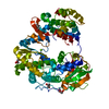
| ||||||||
|---|---|---|---|---|---|---|---|---|---|
| 1 |
| ||||||||
| Unit cell |
|
-Components
| #1: Protein | Mass: 71208.398 Da / Num. of mol.: 1 Source method: isolated from a genetically manipulated source Source: (gene. exp.) Jingmen tick virus / Strain: YN-flaviV / Gene: segment 1 / Production host:  | ||||||
|---|---|---|---|---|---|---|---|
| #2: Chemical | ChemComp-MG /   Mass: 24.305 Da / Num. of mol.: 1 / Source method: obtained synthetically / Formula: Mg Mass: 24.305 Da / Num. of mol.: 1 / Source method: obtained synthetically / Formula: Mg | ||||||
| #3: Chemical | ChemComp-GOL /   Mass: 92.094 Da / Num. of mol.: 4 / Source method: obtained synthetically / Formula: C3H8O3 Mass: 92.094 Da / Num. of mol.: 4 / Source method: obtained synthetically / Formula: C3H8O3#4: Chemical |   Mass: 106.120 Da / Num. of mol.: 2 / Source method: obtained synthetically / Formula: C4H10O3 Mass: 106.120 Da / Num. of mol.: 2 / Source method: obtained synthetically / Formula: C4H10O3#5: Water | ChemComp-HOH / |  Mass: 18.015 Da / Num. of mol.: 419 / Source method: isolated from a natural source / Formula: H2O Mass: 18.015 Da / Num. of mol.: 419 / Source method: isolated from a natural source / Formula: H2OHas ligand of interest | N | |
-Experimental details
-Experiment
| Experiment | Method: X-RAY DIFFRACTION / Number of used crystals: 1 |
|---|
- Sample preparation
| Crystal | Density Matthews: 2.36 Å3/Da / Density % sol: 47.95 % |
|---|---|
| Crystal grow | Temperature: 289 K / Method: vapor diffusion / Details: potassium sodium tartrate tetrahyrate |
-Data collection
| Diffraction | Mean temperature: 100 K / Serial crystal experiment: N | ||||||||||||||||||||||||||||||||||||||||||||||||||||||||||||||||||||||||||||||||||||||||||||||||||||||||||||||
|---|---|---|---|---|---|---|---|---|---|---|---|---|---|---|---|---|---|---|---|---|---|---|---|---|---|---|---|---|---|---|---|---|---|---|---|---|---|---|---|---|---|---|---|---|---|---|---|---|---|---|---|---|---|---|---|---|---|---|---|---|---|---|---|---|---|---|---|---|---|---|---|---|---|---|---|---|---|---|---|---|---|---|---|---|---|---|---|---|---|---|---|---|---|---|---|---|---|---|---|---|---|---|---|---|---|---|---|---|---|---|---|
| Diffraction source | Source: SYNCHROTRON / Site: SSRF / Beamline: BL17U / Wavelength: 0.9792 Å | ||||||||||||||||||||||||||||||||||||||||||||||||||||||||||||||||||||||||||||||||||||||||||||||||||||||||||||||
| Detector | Type: ADSC QUANTUM 315r / Detector: CCD / Date: Dec 7, 2019 | ||||||||||||||||||||||||||||||||||||||||||||||||||||||||||||||||||||||||||||||||||||||||||||||||||||||||||||||
| Radiation | Protocol: SINGLE WAVELENGTH / Monochromatic (M) / Laue (L): M / Scattering type: x-ray | ||||||||||||||||||||||||||||||||||||||||||||||||||||||||||||||||||||||||||||||||||||||||||||||||||||||||||||||
| Radiation wavelength | Wavelength: 0.9792 Å / Relative weight: 1 | ||||||||||||||||||||||||||||||||||||||||||||||||||||||||||||||||||||||||||||||||||||||||||||||||||||||||||||||
| Reflection | Resolution: 1.84→50 Å / Num. obs: 58904 / % possible obs: 99.1 % / Redundancy: 6.7 % / CC1/2: 0.999 / CC star: 1 / Rmerge(I) obs: 0.077 / Rpim(I) all: 0.031 / Rrim(I) all: 0.083 / Χ2: 1.012 / Net I/σ(I): 9.6 / Num. measured all: 395284 | ||||||||||||||||||||||||||||||||||||||||||||||||||||||||||||||||||||||||||||||||||||||||||||||||||||||||||||||
| Reflection shell | Diffraction-ID: 1
|
- Processing
| Software |
| ||||||||||||||||||||||||||||||||||||||||||||||||||||||||||||||||||||||||||||||||||||||||||||||||||||||||||||||||||||||||||||||||||||||||||||||||||||||||||
|---|---|---|---|---|---|---|---|---|---|---|---|---|---|---|---|---|---|---|---|---|---|---|---|---|---|---|---|---|---|---|---|---|---|---|---|---|---|---|---|---|---|---|---|---|---|---|---|---|---|---|---|---|---|---|---|---|---|---|---|---|---|---|---|---|---|---|---|---|---|---|---|---|---|---|---|---|---|---|---|---|---|---|---|---|---|---|---|---|---|---|---|---|---|---|---|---|---|---|---|---|---|---|---|---|---|---|---|---|---|---|---|---|---|---|---|---|---|---|---|---|---|---|---|---|---|---|---|---|---|---|---|---|---|---|---|---|---|---|---|---|---|---|---|---|---|---|---|---|---|---|---|---|---|---|---|
| Refinement | Method to determine structure: MOLECULAR REPLACEMENT Starting model: in-house model determined by SAD Resolution: 1.84→37.11 Å / SU ML: 0.21 / Cross valid method: THROUGHOUT / σ(F): 1.35 / Phase error: 19.75 / Stereochemistry target values: ML
| ||||||||||||||||||||||||||||||||||||||||||||||||||||||||||||||||||||||||||||||||||||||||||||||||||||||||||||||||||||||||||||||||||||||||||||||||||||||||||
| Solvent computation | Shrinkage radii: 0.9 Å / VDW probe radii: 1.11 Å / Solvent model: FLAT BULK SOLVENT MODEL | ||||||||||||||||||||||||||||||||||||||||||||||||||||||||||||||||||||||||||||||||||||||||||||||||||||||||||||||||||||||||||||||||||||||||||||||||||||||||||
| Refinement step | Cycle: LAST / Resolution: 1.84→37.11 Å
| ||||||||||||||||||||||||||||||||||||||||||||||||||||||||||||||||||||||||||||||||||||||||||||||||||||||||||||||||||||||||||||||||||||||||||||||||||||||||||
| Refine LS restraints |
| ||||||||||||||||||||||||||||||||||||||||||||||||||||||||||||||||||||||||||||||||||||||||||||||||||||||||||||||||||||||||||||||||||||||||||||||||||||||||||
| LS refinement shell |
|
