 Movie
Movie Controller


+ Open data

- Basic information
| Entry | Database: PDB / ID: 8wik | ||||||||||||
|---|---|---|---|---|---|---|---|---|---|---|---|---|---|
| Title | Crystal structure of human FSP1 | ||||||||||||
 Components Components | Ferroptosis suppressor protein 1 | ||||||||||||
 Keywords Keywords | OXIDOREDUCTASE / FSP1 / Ferroptosis / FAD / NADH / inhibitor | ||||||||||||
| Function / homology |  Function and homology information Function and homology informationelectron-transferring-flavoprotein dehydrogenase activity / ubiquinone metabolic process / Oxidoreductases; Acting on NADH or NADPH; With a quinone or similar compound as acceptor / vitamin K metabolic process / regulation of cellular response to oxidative stress / oxidoreductase activity, acting on NAD(P)H, quinone or similar compound as acceptor / cellular detoxification / negative regulation of ferroptosis / TP53 Regulates Transcription of Genes Involved in Cytochrome C Release / apoptotic mitochondrial changes ...electron-transferring-flavoprotein dehydrogenase activity / ubiquinone metabolic process / Oxidoreductases; Acting on NADH or NADPH; With a quinone or similar compound as acceptor / vitamin K metabolic process / regulation of cellular response to oxidative stress / oxidoreductase activity, acting on NAD(P)H, quinone or similar compound as acceptor / cellular detoxification / negative regulation of ferroptosis / TP53 Regulates Transcription of Genes Involved in Cytochrome C Release / apoptotic mitochondrial changes / lipid droplet / flavin adenine dinucleotide binding / mitochondrial outer membrane / positive regulation of apoptotic process / mitochondrion / extracellular space / DNA binding / nucleus / plasma membrane / cytosol / cytoplasm Similarity search - Function | ||||||||||||
| Biological species |  Homo sapiens (human) Homo sapiens (human) | ||||||||||||
| Method |  X-RAY DIFFRACTION / SYNCHROTRON / MOLECULAR REPLACEMENT / Resolution: 2 Å X-RAY DIFFRACTION / SYNCHROTRON / MOLECULAR REPLACEMENT / Resolution: 2 Å | ||||||||||||
 Authors Authors | Feng, S. / Huang, X. / Tang, D. / Qi, S. | ||||||||||||
| Funding support |  China, 3items China, 3items
| ||||||||||||
 Citation Citation | Journal: MedComm (2020) / Year: 2024 Title: The crystal structure of human ferroptosis suppressive protein 1 in complex with flavin adenine dinucleotide and nicotinamide adenine nucleotide. Authors: Feng, S. / Huang, X. / Tang, D. / Liu, X. / Ouyang, L. / Yang, D. / Wang, K. / Liao, B. / Qi, S. | ||||||||||||
| History |
|
- Structure visualization
| Structure viewer | Molecule: MolmilJmol/JSmol |
|---|
- Downloads & links
-Download
| PDBx/mmCIF format | 8wik.cif.gz | 196.1 KB | Display | PDBx/mmCIF format |
|---|---|---|---|---|
| PDB format | pdb8wik.ent.gz | 129.7 KB | Display | PDB format |
| PDBx/mmJSON format | 8wik.json.gz | Tree view | PDBx/mmJSON format | |
| Others |  Other downloads Other downloads |
-Validation report
| Summary document | 8wik_validation.pdf.gz | 949.5 KB | Display | wwPDB validaton report |
|---|---|---|---|---|
| Full document | 8wik_full_validation.pdf.gz | 953.2 KB | Display | |
| Data in XML | 8wik_validation.xml.gz | 17.9 KB | Display | |
| Data in CIF | 8wik_validation.cif.gz | 25.5 KB | Display | |
| Arichive directory | https://data.pdbj.org/pub/pdb/validation_reports/wi/8wikftp://data.pdbj.org/pub/pdb/validation_reports/wi/8wik | HTTPS FTP |
-Related structure data
| Similar structure data |
|---|
-Links
 PDBj
PDBj


- Assembly
| Deposited unit | 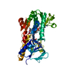
| ||||||||||||
|---|---|---|---|---|---|---|---|---|---|---|---|---|---|
| 1 |
| ||||||||||||
| Unit cell |
|
-Components
| #1: Protein | Mass: 40573.598 Da / Num. of mol.: 1 Source method: isolated from a genetically manipulated source Source: (gene. exp.) Homo sapiens (human) / Gene: AIFM2, AMID, PRG3 / Production host:   Spodoptera frugiperda (fall armyworm) Spodoptera frugiperda (fall armyworm)References: UniProt: Q9BRQ8, Oxidoreductases; Acting on NADH or NADPH; With a quinone or similar compound as acceptor |
|---|---|
| #2: Chemical | ChemComp-6FA / 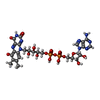  Mass: 801.549 Da / Num. of mol.: 1 / Source method: obtained synthetically / Formula: C27H33N9O16P2 / Feature type: SUBJECT OF INVESTIGATION Mass: 801.549 Da / Num. of mol.: 1 / Source method: obtained synthetically / Formula: C27H33N9O16P2 / Feature type: SUBJECT OF INVESTIGATION |
| #3: Chemical | ChemComp-NAD /   Mass: 663.425 Da / Num. of mol.: 1 / Source method: obtained synthetically / Formula: C21H27N7O14P2 / Feature type: SUBJECT OF INVESTIGATION / Comment: NAD*YM Mass: 663.425 Da / Num. of mol.: 1 / Source method: obtained synthetically / Formula: C21H27N7O14P2 / Feature type: SUBJECT OF INVESTIGATION / Comment: NAD*YM |
| #4: Water | ChemComp-HOH /  Mass: 18.015 Da / Num. of mol.: 197 / Source method: isolated from a natural source / Formula: H2O Mass: 18.015 Da / Num. of mol.: 197 / Source method: isolated from a natural source / Formula: H2O |
| Has ligand of interest | Y |
-Experimental details
-Experiment
| Experiment | Method: X-RAY DIFFRACTION / Number of used crystals: 1 |
|---|
- Sample preparation
| Crystal | Density Matthews: 2.93 Å3/Da / Density % sol: 58.05 % |
|---|---|
| Crystal grow | Temperature: 291 K / Method: vapor diffusion, hanging drop / Details: HEPES , MgCl2, PEG3350 |
-Data collection
| Diffraction | Mean temperature: 100 K / Ambient temp details: 291 / Serial crystal experiment: N | ||||||||||||||||||||||||||||||||||||||||||||||||||||||||||||||||||||||||||||||||||||||||||||||||||||||||||||||
|---|---|---|---|---|---|---|---|---|---|---|---|---|---|---|---|---|---|---|---|---|---|---|---|---|---|---|---|---|---|---|---|---|---|---|---|---|---|---|---|---|---|---|---|---|---|---|---|---|---|---|---|---|---|---|---|---|---|---|---|---|---|---|---|---|---|---|---|---|---|---|---|---|---|---|---|---|---|---|---|---|---|---|---|---|---|---|---|---|---|---|---|---|---|---|---|---|---|---|---|---|---|---|---|---|---|---|---|---|---|---|---|
| Diffraction source | Source: SYNCHROTRON / Site: SSRF / Beamline: BL19U1 / Wavelength: 1 Å | ||||||||||||||||||||||||||||||||||||||||||||||||||||||||||||||||||||||||||||||||||||||||||||||||||||||||||||||
| Detector | Type: MAR CCD 165 mm / Detector: CCD / Date: Jan 15, 2023 | ||||||||||||||||||||||||||||||||||||||||||||||||||||||||||||||||||||||||||||||||||||||||||||||||||||||||||||||
| Radiation | Protocol: SINGLE WAVELENGTH / Monochromatic (M) / Laue (L): M / Scattering type: x-ray | ||||||||||||||||||||||||||||||||||||||||||||||||||||||||||||||||||||||||||||||||||||||||||||||||||||||||||||||
| Radiation wavelength | Wavelength: 1 Å / Relative weight: 1 | ||||||||||||||||||||||||||||||||||||||||||||||||||||||||||||||||||||||||||||||||||||||||||||||||||||||||||||||
| Reflection | Resolution: 2→50 Å / Num. obs: 34829 / % possible obs: 100 % / Redundancy: 9.8 % / Biso Wilson estimate: 24.58 Å2 / CC1/2: 0.969 / CC star: 0.992 / Rmerge(I) obs: 0.118 / Rpim(I) all: 0.04 / Rrim(I) all: 0.125 / Χ2: 0.959 / Net I/σ(I): 4.6 / Num. measured all: 339711 | ||||||||||||||||||||||||||||||||||||||||||||||||||||||||||||||||||||||||||||||||||||||||||||||||||||||||||||||
| Reflection shell | Diffraction-ID: 1
|
- Processing
| Software |
| ||||||||||||||||||||||||||||||||||||||||||||||||||||||||||||||||||||||||||||||||||||||||||||||||||||||||||||||||||||||||||||||||||||||||||||
|---|---|---|---|---|---|---|---|---|---|---|---|---|---|---|---|---|---|---|---|---|---|---|---|---|---|---|---|---|---|---|---|---|---|---|---|---|---|---|---|---|---|---|---|---|---|---|---|---|---|---|---|---|---|---|---|---|---|---|---|---|---|---|---|---|---|---|---|---|---|---|---|---|---|---|---|---|---|---|---|---|---|---|---|---|---|---|---|---|---|---|---|---|---|---|---|---|---|---|---|---|---|---|---|---|---|---|---|---|---|---|---|---|---|---|---|---|---|---|---|---|---|---|---|---|---|---|---|---|---|---|---|---|---|---|---|---|---|---|---|---|---|
| Refinement | Method to determine structure: MOLECULAR REPLACEMENT / Resolution: 2→35.26 Å / SU ML: 0.2611 / Cross valid method: FREE R-VALUE / σ(F): 1.34 / Phase error: 28.0098 Stereochemistry target values: GeoStd + Monomer Library + CDL v1.2
| ||||||||||||||||||||||||||||||||||||||||||||||||||||||||||||||||||||||||||||||||||||||||||||||||||||||||||||||||||||||||||||||||||||||||||||
| Solvent computation | Shrinkage radii: 0.9 Å / VDW probe radii: 1.1 Å / Solvent model: FLAT BULK SOLVENT MODEL | ||||||||||||||||||||||||||||||||||||||||||||||||||||||||||||||||||||||||||||||||||||||||||||||||||||||||||||||||||||||||||||||||||||||||||||
| Displacement parameters | Biso mean: 35.59 Å2 | ||||||||||||||||||||||||||||||||||||||||||||||||||||||||||||||||||||||||||||||||||||||||||||||||||||||||||||||||||||||||||||||||||||||||||||
| Refinement step | Cycle: LAST / Resolution: 2→35.26 Å
| ||||||||||||||||||||||||||||||||||||||||||||||||||||||||||||||||||||||||||||||||||||||||||||||||||||||||||||||||||||||||||||||||||||||||||||
| Refine LS restraints |
| ||||||||||||||||||||||||||||||||||||||||||||||||||||||||||||||||||||||||||||||||||||||||||||||||||||||||||||||||||||||||||||||||||||||||||||
| LS refinement shell |
| ||||||||||||||||||||||||||||||||||||||||||||||||||||||||||||||||||||||||||||||||||||||||||||||||||||||||||||||||||||||||||||||||||||||||||||
| Refinement TLS params. | Method: refined / Origin x: 16.4241703844 Å / Origin y: -9.36275331131 Å / Origin z: -18.5992812359 Å
| ||||||||||||||||||||||||||||||||||||||||||||||||||||||||||||||||||||||||||||||||||||||||||||||||||||||||||||||||||||||||||||||||||||||||||||
| Refinement TLS group | Selection details: (chain 'A' and resid 10 through 402) |
