[English] 日本語
Yorodumi
- PDB-8wct: The crystal structure of the CHASE4 domain of iron-sensetive memb... -

+
Open data


ID or keywords:

Loading...

-
Basic information

Entry
Database: PDB / ID: 8wct
TitleThe crystal structure of the CHASE4 domain of iron-sensetive membrane protein (IsmP,Uniprot ID:Q9I243)
ComponentsBifunctional diguanylate cyclase/phosphodiesterase
KeywordsMEMBRANE PROTEIN / CHEASE4 domain / iron-sensetive
Function / homology
Function and homology information


CHASE4 / CHASE4 domain / Putative diguanylate phosphodiesterase / EAL domain / EAL domain superfamily / EAL domain profile. / EAL domain / Diguanylate cyclase, GGDEF domain / diguanylate cyclase / GGDEF domain profile. ...CHASE4 / CHASE4 domain / Putative diguanylate phosphodiesterase / EAL domain / EAL domain superfamily / EAL domain profile. / EAL domain / Diguanylate cyclase, GGDEF domain / diguanylate cyclase / GGDEF domain profile. / GGDEF domain / Nucleotide cyclase / PAS domain / PAS repeat profile. / PAS domain / PAS domain superfamily / Reverse transcriptase/Diguanylate cyclase domain
Similarity search - Domain/homology
Bifunctional diguanylate cyclase/phosphodiesterase
Similarity search - Component
Biological speciesPseudomonas aeruginosa (bacteria)
MethodX-RAY DIFFRACTION / SYNCHROTRON / MOLECULAR REPLACEMENT / Resolution: 1.9 Å
AuthorsWang, C.C.
Funding support China, 1items
OrganizationGrant numberCountry
National Natural Science Foundation of China (NSFC) China
Citation
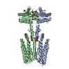
Journal: Nat Commun / Year: 2024
Title: A c-di-GMP signaling module controls responses to iron in Pseudomonas aeruginosa.
Authors: Xueliang Zhan / Kuo Zhang / Chenchen Wang / Qiao Fan / Xiujia Tang / Xi Zhang / Ke Wang / Yang Fu / Haihua Liang /
Abstract: Cyclic dimeric guanosine monophosphate (c-di-GMP) serves as a bacterial second messenger that modulates various processes including biofilm formation, motility, and host-microbe symbiosis. Numerous ...Cyclic dimeric guanosine monophosphate (c-di-GMP) serves as a bacterial second messenger that modulates various processes including biofilm formation, motility, and host-microbe symbiosis. Numerous studies have conducted comprehensive analysis of c-di-GMP. However, the mechanisms by which certain environmental signals such as iron control intracellular c-di-GMP levels are unclear. Here, we show that iron regulates c-di-GMP levels in Pseudomonas aeruginosa by modulating the interaction between an iron-sensing protein, IsmP, and a diguanylate cyclase, ImcA. Binding of iron to the CHASE4 domain of IsmP inhibits the IsmP-ImcA interaction, which leads to increased c-di-GMP synthesis by ImcA, thus promoting biofilm formation and reducing bacterial motility. Structural characterization of the apo-CHASE4 domain and its binding to iron allows us to pinpoint residues defining its specificity. In addition, the cryo-electron microscopy structure of ImcA in complex with a c-di-GMP analog (GMPCPP) suggests a unique conformation in which the compound binds to the catalytic pockets and to the membrane-proximal side located at the cytoplasm. Thus, our results indicate that a CHASE4 domain directly senses iron and modulates the crosstalk between c-di-GMP metabolic enzymes.
History
DepositionSep 13, 2023Deposition site: PDBJ / Processing site: PDBJ
Revision 1.0Feb 14, 2024Provider: repository / Type: Initial release
Revision 1.1Mar 13, 2024Group: Database references / Category: citation / citation_author
Item: _citation.country / _citation.journal_abbrev ..._citation.country / _citation.journal_abbrev / _citation.journal_id_CSD / _citation.journal_id_ISSN / _citation.journal_volume / _citation.page_first / _citation.page_last / _citation.pdbx_database_id_DOI / _citation.pdbx_database_id_PubMed / _citation.title / _citation.year

-
Structure visualization

Structure viewerMolecule:
MolmilJmol/JSmol

Downloads & links

-
Assembly

Deposited unit
A: Bifunctional diguanylate cyclase/phosphodiesterase
B: Bifunctional diguanylate cyclase/phosphodiesterase
hetero molecules


Theoretical massNumber of molelcules
Total (without water)54,3423
Polymers54,2502
Non-polymers921
Water6,990388
1


  • Idetical with deposited unit
  • defined by author&software
  • Evidence: gel filtration
TypeNameSymmetry operationNumber
identity operation1_555x,y,z1
Unit cell
Length a, b, c (Å)64.431, 75.205, 104.080
Angle α, β, γ (deg.)90.000, 90.000, 90.000
Int Tables number18
Space group name H-MP22121
Space group name HallP22ab(z,x,y)

-
Components

#1: Protein Bifunctional diguanylate cyclase/phosphodiesterase / Iron-sensetive membrane protein


Mass: 27125.186 Da / Num. of mol.: 2
Source method: isolated from a genetically manipulated source
Source: (gene. exp.) Pseudomonas aeruginosa (bacteria) / Gene: PA2072 / Production host: Escherichia coli (E. coli) / References: UniProt: Q9I243
#2: Chemical ChemComp-GOL / GLYCEROL / GLYCERIN / PROPANE-1,2,3-TRIOL


Mass: 92.094 Da / Num. of mol.: 1 / Source method: obtained synthetically / Formula: C3H8O3
#3: Water ChemComp-HOH / water


Mass: 18.015 Da / Num. of mol.: 388 / Source method: isolated from a natural source / Formula: H2O
Has ligand of interestN

-
Experimental details

-
Experiment

ExperimentMethod: X-RAY DIFFRACTION / Number of used crystals: 1

-
Sample preparation

CrystalDensity Matthews: 2.32 Å3/Da / Density % sol: 47.08 %
Crystal growTemperature: 289 K / Method: vapor diffusion, sitting drop
Details: 0.2 M sodium acetate lithium, 20% PEG3350, 0.1 M Bis-Tris propane, pH 7.9

-
Data collection

DiffractionMean temperature: 100 K / Serial crystal experiment: N
Diffraction sourceSource: SYNCHROTRON / Site: SSRF / Beamline: BL18U1 / Wavelength: 0.97915 Å
DetectorType: DECTRIS PILATUS 6M / Detector: PIXEL / Date: Apr 1, 2023
RadiationProtocol: SINGLE WAVELENGTH / Monochromatic (M) / Laue (L): M / Scattering type: x-ray
Radiation wavelengthWavelength: 0.97915 Å / Relative weight: 1
ReflectionResolution: 1.9→50 Å / Num. obs: 40453 / % possible obs: 99.9 % / Redundancy: 11.4 % / Biso Wilson estimate: 23.24 Å2 / CC1/2: 0.998 / Rmerge(I) obs: 0.119 / Rrim(I) all: 0.125 / Net I/σ(I): 13.84
Reflection shell
Resolution (Å)Rmerge(I) obsNum. unique obsCC1/2Rrim(I) allDiffraction-ID
1.9-2.020.55464750.980.581
2.02-2.150.3960860.9870.4091
2.15-2.330.28356500.9920.2971
2.33-2.550.21452390.9950.2241
2.55-2.850.15547630.9970.1621
2.85-3.290.10942230.9980.1151
3.29-4.020.07836070.9980.0821
4.02-5.670.06428370.9980.0671
1.9-1.950.0616760.9980.0641

-
Processing

Software
NameVersionClassification
PHENIXdev_4788model building
PHENIXdev_4788refinement
Aimlessdata scaling
XDSdata reduction
PHASERphasing
RefinementMethod to determine structure: MOLECULAR REPLACEMENT / Resolution: 1.9→42.79 Å / SU ML: 0.2299 / Cross valid method: FREE R-VALUE / σ(F): 1.34 / Phase error: 32.1576
Stereochemistry target values: GeoStd + Monomer Library + CDL v1.2
RfactorNum. reflection% reflection
Rfree0.2634 1973 4.88 %
Rwork0.2252 38480 -
obs0.2271 40453 99.69 %
Solvent computationShrinkage radii: 0.9 Å / VDW probe radii: 1.1 Å / Solvent model: FLAT BULK SOLVENT MODEL
Displacement parametersBiso mean: 29.72 Å2
Refinement stepCycle: LAST / Resolution: 1.9→42.79 Å
ProteinNucleic acidLigandSolventTotal
Num. atoms3276 0 6 388 3670
Refine LS restraints
Refine-IDTypeDev idealNumber
X-RAY DIFFRACTIONf_bond_d0.00663345
X-RAY DIFFRACTIONf_angle_d0.94294537
X-RAY DIFFRACTIONf_chiral_restr0.0544500
X-RAY DIFFRACTIONf_plane_restr0.0074598
X-RAY DIFFRACTIONf_dihedral_angle_d12.06591192
LS refinement shell
Resolution (Å)Rfactor RfreeNum. reflection RfreeRfactor RworkNum. reflection RworkRefine-ID% reflection obs (%)
1.9-1.950.37531480.28552695X-RAY DIFFRACTION99.13
1.95-20.29661290.25082707X-RAY DIFFRACTION99.72
2-2.060.27811380.23782701X-RAY DIFFRACTION99.68
2.06-2.130.25611270.23392712X-RAY DIFFRACTION99.89
2.13-2.20.3081510.22652718X-RAY DIFFRACTION99.62
2.2-2.290.29191310.2272714X-RAY DIFFRACTION99.68
2.29-2.390.26431100.22362770X-RAY DIFFRACTION99.62
2.39-2.520.2771320.23252736X-RAY DIFFRACTION99.69
2.52-2.680.25421500.23892745X-RAY DIFFRACTION99.79
2.68-2.880.2711190.22842747X-RAY DIFFRACTION99.79
2.88-3.170.30611550.2262750X-RAY DIFFRACTION99.93
3.18-3.630.20741540.21352764X-RAY DIFFRACTION99.83
3.63-4.580.25071590.20742798X-RAY DIFFRACTION99.66
4.58-42.790.25861700.22312923X-RAY DIFFRACTION99.68

+
About Yorodumi

-
News

-
Feb 9, 2022. New format data for meta-information of EMDB entries

New format data for meta-information of EMDB entries

  • Version 3 of the EMDB header file is now the official format.
  • The previous official version 1.9 will be removed from the archive.

Related info.:EMDB header

External links:wwPDB to switch to version 3 of the EMDB data model

-
Aug 12, 2020. Covid-19 info

Covid-19 info

URL: https://pdbjlvh1.pdbj.org/emnavi/covid19.php

New page: Covid-19 featured information page in EM Navigator.

Related info.:Covid-19 info / Mar 5, 2020. Novel coronavirus structure data

+
Mar 5, 2020. Novel coronavirus structure data

Novel coronavirus structure data

Related info.:Yorodumi Speices / Aug 12, 2020. Covid-19 info

External links:COVID-19 featured content - PDBj / Molecule of the Month (242):Coronavirus Proteases

+
Jan 31, 2019. EMDB accession codes are about to change! (news from PDBe EMDB page)

EMDB accession codes are about to change! (news from PDBe EMDB page)

  • The allocation of 4 digits for EMDB accession codes will soon come to an end. Whilst these codes will remain in use, new EMDB accession codes will include an additional digit and will expand incrementally as the available range of codes is exhausted. The current 4-digit format prefixed with “EMD-” (i.e. EMD-XXXX) will advance to a 5-digit format (i.e. EMD-XXXXX), and so on. It is currently estimated that the 4-digit codes will be depleted around Spring 2019, at which point the 5-digit format will come into force.
  • The EM Navigator/Yorodumi systems omit the EMD- prefix.

Related info.:Q: What is EMD? / ID/Accession-code notation in Yorodumi/EM Navigator

External links:EMDB Accession Codes are Changing Soon! / Contact to PDBj

+
Jul 12, 2017. Major update of PDB

Major update of PDB

  • wwPDB released updated PDB data conforming to the new PDBx/mmCIF dictionary.
  • This is a major update changing the version number from 4 to 5, and with Remediation, in which all the entries are updated.
  • In this update, many items about electron microscopy experimental information are reorganized (e.g. em_software).
  • Now, EM Navigator and Yorodumi are based on the updated data.

External links:wwPDB Remediation / Enriched Model Files Conforming to OneDep Data Standards Now Available in the PDB FTP Archive

-
Yorodumi

Thousand views of thousand structures

  • Yorodumi is a browser for structure data from EMDB, PDB, SASBDB, etc.
  • This page is also the successor to EM Navigator detail page, and also detail information page/front-end page for Omokage search.
  • The word "yorodu" (or yorozu) is an old Japanese word meaning "ten thousand". "mi" (miru) is to see.

Related info.:EMDB / PDB / SASBDB / Comparison of 3 databanks / Yorodumi Search / Aug 31, 2016. New EM Navigator & Yorodumi / Yorodumi Papers / Jmol/JSmol / Function and homology information / Changes in new EM Navigator and Yorodumi

Read more