 Movie
Movie Controller

[English] 日本語
 Yorodumi
- PDB-8wbv: The crystal structure of linear mannose with mutant H247F of the ... -
+ Open data

- Basic information
| Entry | Database: PDB / ID: 8wbv | ||||||
|---|---|---|---|---|---|---|---|
| Title | The crystal structure of linear mannose with mutant H247F of the cellobiose 2-epimerase from Caldicellulosiruptor saccharolyticus | ||||||
 Components Components | Cellobiose 2-epimerase | ||||||
 Keywords Keywords | ISOMERASE / cellobiose 2-epimerase / BIOSYNTHETIC PROTEIN | ||||||
| Function / homology |  Function and homology information Function and homology informationcellobiose epimerase / cellobiose epimerase activity / carbohydrate metabolic process Similarity search - Function | ||||||
| Biological species |   Caldicellulosiruptor saccharolyticus (bacteria) Caldicellulosiruptor saccharolyticus (bacteria) | ||||||
| Method |  X-RAY DIFFRACTION / SYNCHROTRON / MOLECULAR REPLACEMENT / Resolution: 1.95 Å X-RAY DIFFRACTION / SYNCHROTRON / MOLECULAR REPLACEMENT / Resolution: 1.95 Å | ||||||
 Authors Authors | Yang, R.J. / Feng, Y.H. | ||||||
| Funding support |  China, 1items China, 1items
| ||||||
 Citation Citation | Journal: Int.J.Biol.Macromol. / Year: 2023 Title: A precise swaying map for how promiscuous cellobiose-2-epimerase operate bi-reaction. Authors: Feng, Y. / Lyu, X. / Cong, Y. / Miao, T. / Fang, B. / Zhang, C. / Shen, Q. / Matthews, M. / Fisher, A.J. / Zhang, J.Z.H. / Zhang, L. / Yang, R. | ||||||
| History |
|
- Structure visualization
| Structure viewer | Molecule: MolmilJmol/JSmol |
|---|
- Downloads & links
-Download
| PDBx/mmCIF format | 8wbv.cif.gz | 183.4 KB | Display | PDBx/mmCIF format |
|---|---|---|---|---|
| PDB format | pdb8wbv.ent.gz | 143.4 KB | Display | PDB format |
| PDBx/mmJSON format | 8wbv.json.gz | Tree view | PDBx/mmJSON format | |
| Others |  Other downloads Other downloads |
-Validation report
| Arichive directory | https://data.pdbj.org/pub/pdb/validation_reports/wb/8wbvftp://data.pdbj.org/pub/pdb/validation_reports/wb/8wbv | HTTPS FTP |
|---|
-Related structure data

-Links
 PDBj
PDBj
- Assembly
| Deposited unit | 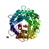
| ||||||||
|---|---|---|---|---|---|---|---|---|---|
| 1 |
| ||||||||
| Unit cell |
|
-Components
| #1: Protein | Mass: 46623.121 Da / Num. of mol.: 1 / Mutation: H247F Source method: isolated from a genetically manipulated source Source: (gene. exp.) Caldicellulosiruptor saccharolyticus (bacteria)Strain: ATCC 43494 / DSM 8903 / Tp8T 6331 / Gene: Csac_0294 / Production host: |
|---|---|
| #2: Sugar | ChemComp-DNO /   Type: D-saccharide / Mass: 180.156 Da / Num. of mol.: 1 / Source method: obtained synthetically / Formula: C6H12O6 / Feature type: SUBJECT OF INVESTIGATION Type: D-saccharide / Mass: 180.156 Da / Num. of mol.: 1 / Source method: obtained synthetically / Formula: C6H12O6 / Feature type: SUBJECT OF INVESTIGATION |
| #3: Sugar | ChemComp-BMA /   Type: D-saccharide, beta linking / Mass: 180.156 Da / Num. of mol.: 1 / Source method: obtained synthetically / Formula: C6H12O6 Type: D-saccharide, beta linking / Mass: 180.156 Da / Num. of mol.: 1 / Source method: obtained synthetically / Formula: C6H12O6 |
| #4: Chemical | ChemComp-EDO /   Mass: 62.068 Da / Num. of mol.: 1 / Source method: obtained synthetically / Formula: C2H6O2 Mass: 62.068 Da / Num. of mol.: 1 / Source method: obtained synthetically / Formula: C2H6O2 |
| #5: Water | ChemComp-HOH /  Mass: 18.015 Da / Num. of mol.: 128 / Source method: isolated from a natural source / Formula: H2O Mass: 18.015 Da / Num. of mol.: 128 / Source method: isolated from a natural source / Formula: H2O |
| Has ligand of interest | Y |
| Has protein modification | N |
-Experimental details
-Experiment
| Experiment | Method: X-RAY DIFFRACTION / Number of used crystals: 1 |
|---|
- Sample preparation
| Crystal | Density Matthews: 2.2 Å3/Da / Density % sol: 44.12 % |
|---|---|
| Crystal grow | Temperature: 293 K / Method: vapor diffusion, hanging drop / pH: 8.5 / Details: 0.8M NaCl,0.1Tris-HCI pH 8.5, 32% PEG3350 |
-Data collection
| Diffraction | Mean temperature: 293 K / Serial crystal experiment: N |
|---|---|
| Diffraction source | Source: SYNCHROTRON / Site: SSRL  / Beamline: BL12-2 / Wavelength: 0.9795 Å / Beamline: BL12-2 / Wavelength: 0.9795 Å |
| Detector | Type: DECTRIS PILATUS3 6M / Detector: PIXEL / Date: Feb 15, 2017 |
| Radiation | Protocol: SINGLE WAVELENGTH / Monochromatic (M) / Laue (L): M / Scattering type: x-ray |
| Radiation wavelength | Wavelength: 0.9795 Å / Relative weight: 1 |
| Reflection | Resolution: 1.95→50 Å / Num. obs: 28308 / % possible obs: 92 % / Observed criterion σ(I): -3 / Redundancy: 3.27 % / CC1/2: 0.995 / Rmerge(I) obs: 0.084 / Net I/σ(I): 11.35 |
| Reflection shell | Resolution: 1.95→2 Å / Redundancy: 3.62 % / Rmerge(I) obs: 0.521 / Mean I/σ(I) obs: 2.44 / Num. unique obs: 2179 / CC1/2: 0.807 / % possible all: 96.6 |
- Processing
| Software |
| |||||||||||||||||||||||||||||||||||||||||||||||||||||||||||||||||||||||||||||
|---|---|---|---|---|---|---|---|---|---|---|---|---|---|---|---|---|---|---|---|---|---|---|---|---|---|---|---|---|---|---|---|---|---|---|---|---|---|---|---|---|---|---|---|---|---|---|---|---|---|---|---|---|---|---|---|---|---|---|---|---|---|---|---|---|---|---|---|---|---|---|---|---|---|---|---|---|---|---|
| Refinement | Method to determine structure: MOLECULAR REPLACEMENT / Resolution: 1.95→29.877 Å / SU ML: 0.23 / Cross valid method: NONE / σ(F): 1.34 / Phase error: 21.72 / Stereochemistry target values: ML
| |||||||||||||||||||||||||||||||||||||||||||||||||||||||||||||||||||||||||||||
| Solvent computation | Shrinkage radii: 0.9 Å / VDW probe radii: 1.11 Å / Solvent model: FLAT BULK SOLVENT MODEL | |||||||||||||||||||||||||||||||||||||||||||||||||||||||||||||||||||||||||||||
| Refinement step | Cycle: LAST / Resolution: 1.95→29.877 Å
| |||||||||||||||||||||||||||||||||||||||||||||||||||||||||||||||||||||||||||||
| Refine LS restraints |
| |||||||||||||||||||||||||||||||||||||||||||||||||||||||||||||||||||||||||||||
| LS refinement shell |
| |||||||||||||||||||||||||||||||||||||||||||||||||||||||||||||||||||||||||||||
| Refinement TLS params. | Method: refined / Origin x: 27.0411 Å / Origin y: 1.0133 Å / Origin z: -9.3553 Å
| |||||||||||||||||||||||||||||||||||||||||||||||||||||||||||||||||||||||||||||
| Refinement TLS group | Selection details: all |
