 Movie
Movie Controller

[English] 日本語
 Yorodumi
- PDB-8w91: Azumapecten Farreri homopolymeric ferritin mutant - H2KE exposed ... -
+ Open data

- Basic information
| Entry | Database: PDB / ID: 8w91 | ||||||
|---|---|---|---|---|---|---|---|
| Title | Azumapecten Farreri homopolymeric ferritin mutant - H2KE exposed to H2O2 for 3 min | ||||||
 Components Components | Ferritin | ||||||
 Keywords Keywords | METAL BINDING PROTEIN / enzyme | ||||||
| Function / homology |  Function and homology information Function and homology informationferroxidase / ferroxidase activity / ferric iron binding / iron ion transport / ferrous iron binding / intracellular iron ion homeostasis / cytoplasm Similarity search - Function | ||||||
| Biological species |  Azumapecten farreri (Farrer's scallop) Azumapecten farreri (Farrer's scallop) | ||||||
| Method |  X-RAY DIFFRACTION / SYNCHROTRON / MOLECULAR REPLACEMENT / Resolution: 2.12 Å X-RAY DIFFRACTION / SYNCHROTRON / MOLECULAR REPLACEMENT / Resolution: 2.12 Å | ||||||
 Authors Authors | Zhang, T. / Jiao, R. | ||||||
| Funding support |  China, 1items China, 1items
| ||||||
 Citation Citation | Journal: Inorg.Chem. / Year: 2024 Title: Structural Insights into the Reaction between Hydrogen Peroxide and Di-iron Complexes at the Ferroxidase Center of Ferritin. Authors: Jiao, R. / Zhao, G. / Zhang, T. | ||||||
| History |
|
- Structure visualization
| Structure viewer | Molecule: MolmilJmol/JSmol |
|---|
- Downloads & links
-Download
| PDBx/mmCIF format | 8w91.cif.gz | 54.3 KB | Display | PDBx/mmCIF format |
|---|---|---|---|---|
| PDB format | pdb8w91.ent.gz | 37.9 KB | Display | PDB format |
| PDBx/mmJSON format | 8w91.json.gz | Tree view | PDBx/mmJSON format | |
| Others |  Other downloads Other downloads |
-Validation report
| Arichive directory | https://data.pdbj.org/pub/pdb/validation_reports/w9/8w91ftp://data.pdbj.org/pub/pdb/validation_reports/w9/8w91 | HTTPS FTP |
|---|
-Related structure data


-Links
 PDBj
PDBj


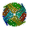
- Assembly
| Deposited unit | 
| |||||||||||||||
|---|---|---|---|---|---|---|---|---|---|---|---|---|---|---|---|---|
| 1 | x 24
| |||||||||||||||
| Unit cell |
| |||||||||||||||
| Components on special symmetry positions |
|
-Components
| #1: Protein | Mass: 19960.330 Da / Num. of mol.: 1 / Mutation: H10K,H121E Source method: isolated from a genetically manipulated source Source: (gene. exp.) Azumapecten farreri (Farrer's scallop) / Production host:  | ||||||
|---|---|---|---|---|---|---|---|
| #2: Chemical |   Mass: 55.845 Da / Num. of mol.: 3 / Source method: obtained synthetically / Formula: Fe / Feature type: SUBJECT OF INVESTIGATION Mass: 55.845 Da / Num. of mol.: 3 / Source method: obtained synthetically / Formula: Fe / Feature type: SUBJECT OF INVESTIGATION#3: Water | ChemComp-HOH / |  Mass: 18.015 Da / Num. of mol.: 186 / Source method: isolated from a natural source / Formula: H2O Mass: 18.015 Da / Num. of mol.: 186 / Source method: isolated from a natural source / Formula: H2OHas ligand of interest | Y | Has protein modification | N | |
-Experimental details
-Experiment
| Experiment | Method: X-RAY DIFFRACTION / Number of used crystals: 1 |
|---|
- Sample preparation
| Crystal | Density Matthews: 3.79 Å3/Da / Density % sol: 67.55 % |
|---|---|
| Crystal grow | Temperature: 293 K / Method: vapor diffusion, hanging drop / Details: 100 mM MOPS, pH 7, 500 mM NaCl |
-Data collection
| Diffraction | Mean temperature: 100 K / Serial crystal experiment: N |
|---|---|
| Diffraction source | Source: SYNCHROTRON / Site: SSRF / Beamline: BL02U1 / Wavelength: 1.6314 Å |
| Detector | Type: DECTRIS EIGER2 S 9M / Detector: PIXEL / Date: Jun 1, 2023 |
| Radiation | Protocol: SINGLE WAVELENGTH / Monochromatic (M) / Laue (L): M / Scattering type: x-ray |
| Radiation wavelength | Wavelength: 1.6314 Å / Relative weight: 1 |
| Reflection | Resolution: 2.12→48.61 Å / Num. obs: 16848 / % possible obs: 94.28 % / Redundancy: 23.8 % / CC1/2: 0.999 / Net I/σ(I): 21.4 |
| Reflection shell | Resolution: 2.122→2.198 Å / Num. unique obs: 1176 / CC1/2: 0.669 |
- Processing
| Software |
| ||||||||||||||||||||||||||||||||||||||||||||||||||||||||||||||||||||||||||||||||||||
|---|---|---|---|---|---|---|---|---|---|---|---|---|---|---|---|---|---|---|---|---|---|---|---|---|---|---|---|---|---|---|---|---|---|---|---|---|---|---|---|---|---|---|---|---|---|---|---|---|---|---|---|---|---|---|---|---|---|---|---|---|---|---|---|---|---|---|---|---|---|---|---|---|---|---|---|---|---|---|---|---|---|---|---|---|---|
| Refinement | Method to determine structure: MOLECULAR REPLACEMENT / Resolution: 2.12→48.61 Å / SU ML: 0.21 / Cross valid method: FREE R-VALUE / σ(F): 1.34 / Phase error: 19.2 / Stereochemistry target values: ML
| ||||||||||||||||||||||||||||||||||||||||||||||||||||||||||||||||||||||||||||||||||||
| Solvent computation | Shrinkage radii: 0.9 Å / VDW probe radii: 1.1 Å / Solvent model: FLAT BULK SOLVENT MODEL | ||||||||||||||||||||||||||||||||||||||||||||||||||||||||||||||||||||||||||||||||||||
| Refinement step | Cycle: LAST / Resolution: 2.12→48.61 Å
| ||||||||||||||||||||||||||||||||||||||||||||||||||||||||||||||||||||||||||||||||||||
| Refine LS restraints |
| ||||||||||||||||||||||||||||||||||||||||||||||||||||||||||||||||||||||||||||||||||||
| LS refinement shell |
|
