 Movie
Movie Controller

[English] 日本語
 Yorodumi
- PDB-8w5z: Crystal structure of tick tyrosylprotein sulfotransferase reveals... -
+ Open data

- Basic information
| Entry | Database: PDB / ID: 8w5z | ||||||
|---|---|---|---|---|---|---|---|
| Title | Crystal structure of tick tyrosylprotein sulfotransferase reveals the activation mechanism of tick anticoagulant protein madanin | ||||||
 Components Components |
| ||||||
 Keywords Keywords | TRANSFERASE / sulfotransferase / sulfation | ||||||
| Function / homology | protein-tyrosine sulfotransferase / protein-tyrosine sulfotransferase activity / Protein-tyrosine sulfotransferase / Sulfotransferase family / P-loop containing nucleoside triphosphate hydrolase / 3-PYRIDINIUM-1-YLPROPANE-1-SULFONATE / ADENOSINE-3'-5'-DIPHOSPHATE / Protein-tyrosine sulfotransferase Function and homology information Function and homology information | ||||||
| Biological species |  Ixodes (arthropod)longicornis species complex (fry) Ixodes (arthropod)longicornis species complex (fry) | ||||||
| Method |  X-RAY DIFFRACTION / SYNCHROTRON / MOLECULAR REPLACEMENT / Resolution: 1.55 Å X-RAY DIFFRACTION / SYNCHROTRON / MOLECULAR REPLACEMENT / Resolution: 1.55 Å | ||||||
 Authors Authors | Yoshimura, M. / Teramoto, T. / Nishimoto, E. / Kakuta, Y. | ||||||
| Funding support |  Japan, 1items Japan, 1items
| ||||||
 Citation Citation | Journal: J.Biol.Chem. / Year: 2024 Title: Crystal structure of tick tyrosylprotein sulfotransferase reveals the activation mechanism of the tick anticoagulant protein madanin. Authors: Yoshimura, M. / Teramoto, T. / Asano, H. / Iwamoto, Y. / Kondo, M. / Nishimoto, E. / Kakuta, Y. | ||||||
| History |
|
- Structure visualization
| Structure viewer | Molecule: MolmilJmol/JSmol |
|---|
- Downloads & links
-Download
| PDBx/mmCIF format | 8w5z.cif.gz | 175.2 KB | Display | PDBx/mmCIF format |
|---|---|---|---|---|
| PDB format | pdb8w5z.ent.gz | 136.6 KB | Display | PDB format |
| PDBx/mmJSON format | 8w5z.json.gz | Tree view | PDBx/mmJSON format | |
| Others |  Other downloads Other downloads |
-Validation report
| Arichive directory | https://data.pdbj.org/pub/pdb/validation_reports/w5/8w5zftp://data.pdbj.org/pub/pdb/validation_reports/w5/8w5z | HTTPS FTP |
|---|
-Related structure data
| Similar structure data |
|---|
-Links
 PDBj
PDBj
- Assembly
| Deposited unit | 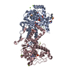
| ||||||||||||
|---|---|---|---|---|---|---|---|---|---|---|---|---|---|
| 1 |
| ||||||||||||
| Unit cell |
| ||||||||||||
| Components on special symmetry positions |
|
-Components
-Protein / Protein/peptide , 2 types, 4 molecules ACBD
| #1: Protein | Mass: 44801.168 Da / Num. of mol.: 2 Source method: isolated from a genetically manipulated source Source: (gene. exp.) Ixodes (arthropod) / Production host:  #2: Protein/peptide | Mass: 1320.227 Da / Num. of mol.: 2 / Source method: obtained synthetically / Source: (synth.) longicornis species complex (fry) |
|---|
-Non-polymers , 5 types, 498 molecules 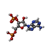
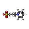


| #3: Chemical |  Type: RNA linking / Mass: 427.201 Da / Num. of mol.: 2 / Source method: obtained synthetically / Formula: C10H15N5O10P2 / Feature type: SUBJECT OF INVESTIGATION Type: RNA linking / Mass: 427.201 Da / Num. of mol.: 2 / Source method: obtained synthetically / Formula: C10H15N5O10P2 / Feature type: SUBJECT OF INVESTIGATION#4: Chemical | ChemComp-1PS /  Mass: 201.243 Da / Num. of mol.: 4 / Source method: isolated from a natural source / Formula: C8H11NO3S / Feature type: SUBJECT OF INVESTIGATION Mass: 201.243 Da / Num. of mol.: 4 / Source method: isolated from a natural source / Formula: C8H11NO3S / Feature type: SUBJECT OF INVESTIGATION#5: Chemical | ChemComp-TRS /  Mass: 122.143 Da / Num. of mol.: 4 / Source method: obtained synthetically / Formula: C4H12NO3 / Feature type: SUBJECT OF INVESTIGATION / Comment: pH buffer*YM Mass: 122.143 Da / Num. of mol.: 4 / Source method: obtained synthetically / Formula: C4H12NO3 / Feature type: SUBJECT OF INVESTIGATION / Comment: pH buffer*YM#6: Chemical | ChemComp-MG /  Mass: 24.305 Da / Num. of mol.: 4 / Source method: isolated from a natural source / Formula: Mg Mass: 24.305 Da / Num. of mol.: 4 / Source method: isolated from a natural source / Formula: Mg#7: Water | ChemComp-HOH / | Mass: 18.015 Da / Num. of mol.: 484 / Source method: isolated from a natural source / Formula: H2O |
|---|
-Details
| Has ligand of interest | Y |
|---|---|
| Has protein modification | Y |
-Experimental details
-Experiment
| Experiment | Method: X-RAY DIFFRACTION / Number of used crystals: 1 |
|---|
- Sample preparation
| Crystal | Density Matthews: 2.81 Å3/Da / Density % sol: 56.28 % |
|---|---|
| Crystal grow | Temperature: 293 K / Method: vapor diffusion, hanging drop / pH: 8 Details: 0.1 M Tris HCl [pH 8.0, 0.1 M magnesium chloride, 15% v/v PEG 400, 16% w/v PEG 8000 |
-Data collection
| Diffraction | Mean temperature: 100 K / Serial crystal experiment: N |
|---|---|
| Diffraction source | Source: SYNCHROTRON / Site: SPring-8 / Beamline: BL45XU / Wavelength: 1 Å |
| Detector | Type: DECTRIS PILATUS 6M / Detector: PIXEL / Date: Dec 12, 2021 |
| Radiation | Protocol: SINGLE WAVELENGTH / Monochromatic (M) / Laue (L): M / Scattering type: x-ray |
| Radiation wavelength | Wavelength: 1 Å / Relative weight: 1 |
| Reflection | Resolution: 1.55→105.5 Å / Num. obs: 150031 / % possible obs: 100 % / Redundancy: 8.8 % / CC1/2: 0.993 / Net I/σ(I): 6.43 |
| Reflection shell | Resolution: 1.55→1.65 Å / Redundancy: 8.6 % / Num. unique obs: 15003 / CC1/2: 0.529 / % possible all: 99.8 |
- Processing
| Software |
| |||||||||||||||||||||||||||||||||||||||||||||||||||||||||||||||||||||||||||||||||||||||||||||||||||||||||
|---|---|---|---|---|---|---|---|---|---|---|---|---|---|---|---|---|---|---|---|---|---|---|---|---|---|---|---|---|---|---|---|---|---|---|---|---|---|---|---|---|---|---|---|---|---|---|---|---|---|---|---|---|---|---|---|---|---|---|---|---|---|---|---|---|---|---|---|---|---|---|---|---|---|---|---|---|---|---|---|---|---|---|---|---|---|---|---|---|---|---|---|---|---|---|---|---|---|---|---|---|---|---|---|---|---|---|
| Refinement | Method to determine structure: MOLECULAR REPLACEMENT / Resolution: 1.55→47.68 Å / SU ML: 0.18 / Cross valid method: FREE R-VALUE / σ(F): 1.34 / Phase error: 20.63 / Stereochemistry target values: ML
| |||||||||||||||||||||||||||||||||||||||||||||||||||||||||||||||||||||||||||||||||||||||||||||||||||||||||
| Solvent computation | Shrinkage radii: 0.9 Å / VDW probe radii: 1.1 Å / Solvent model: FLAT BULK SOLVENT MODEL | |||||||||||||||||||||||||||||||||||||||||||||||||||||||||||||||||||||||||||||||||||||||||||||||||||||||||
| Refinement step | Cycle: LAST / Resolution: 1.55→47.68 Å
| |||||||||||||||||||||||||||||||||||||||||||||||||||||||||||||||||||||||||||||||||||||||||||||||||||||||||
| Refine LS restraints |
| |||||||||||||||||||||||||||||||||||||||||||||||||||||||||||||||||||||||||||||||||||||||||||||||||||||||||
| LS refinement shell |
|
