National Institutes of Health/National Institute of General Medical Sciences (NIH/NIGMS)
R35 GM122462
United States
Citation
Journal: Mol Cell / Year: 2025 Title: Mechanisms of assembly and function of the Hsp70-Hsp40 chaperone machinery. Authors: Yajun Jiang / Ziad Ibrahim / Youlin Xia / Mary Clay / Alexander Myasnikov / Kalyan Immadisetty / Zhilian Xia / Liang Tang / Paolo Rossi / Pritha Ganguly / Jiangshu Liu / Darcie Miller / ...Authors: Yajun Jiang / Ziad Ibrahim / Youlin Xia / Mary Clay / Alexander Myasnikov / Kalyan Immadisetty / Zhilian Xia / Liang Tang / Paolo Rossi / Pritha Ganguly / Jiangshu Liu / Darcie Miller / Meixia Che / Santiago M Palacios / Günter Kramer / Bernd Bukau / Charalampos G Kalodimos / Abstract: Hsp70 and Hsp40 molecular chaperones form a central machinery that remodels client proteins involved in numerous biological processes. Here, we integrated cryo-electron microscopy and nuclear ...Hsp70 and Hsp40 molecular chaperones form a central machinery that remodels client proteins involved in numerous biological processes. Here, we integrated cryo-electron microscopy and nuclear magnetic resonance spectroscopy to determine the architecture of the full-length Hsp70-Hsp40 machinery. The structure of the complex in a physiologically inhibited state reveals distinct regulatory mechanisms. In the active state, the Hsp40 glycine-phenylalanine (G/F)-rich region acts as a pseudo-substrate for Hsp70, directly modulating refolding. This region also maintains Hsp40 in an autoinhibited state; upon binding to Hsp70, the inhibition is disrupted, exposing a cryptic client-binding site that enables client engagement and refolding. Transitions between these states are central to controlling refolding efficiency. Disrupting either the autoinhibited state or the G/F-Hsp70 interaction impairs function and elicits a compensatory heat shock response in cells. Our findings uncover the regulatory dynamics of a fundamental chaperone system, with broad implications for understanding protein homeostasis and the cellular response to stress.
Mass: 18.015 Da / Num. of mol.: 29 / Source method: isolated from a natural source / Formula: H2O
Has ligand of interest
Y
Has protein modification
N
-
Experimental details
-
Experiment
Experiment
Method: X-RAY DIFFRACTION / Number of used crystals: 1
-
Sample preparation
Crystal
Density Matthews: 3.3 Å3/Da / Density % sol: 62.69 %
Crystal grow
Temperature: 293.15 K / Method: vapor diffusion, sitting drop / pH: 6.5 Details: 1:1 volume ratio of protein to reservoir Protein sample: 1.22 mM DnaK, 1.5 mM ADP Protein buffer: 20 mM HEPES pH 7.0, 75mM KCL, 5mM MgSO4 Well solution: 0.1 M NaCacodylate pH 6.5, 0.15 M CaAc, 42% PEG 600
-
Data collection
Diffraction
Mean temperature: 100 K / Serial crystal experiment: N
 Movie
Movie Controller

 Yorodumi
 Open data
 Basic information
 Components
 Keywords
 Function and homology information
 Thermus thermophilus (bacteria)
 X-RAY DIFFRACTION /
  Authors
 United States, 1items
  Citation


 Structure visualization
 Downloads & links
 Other downloads


 PDBj
PDBj
 Assembly

 Escherichia coli BL21(DE3) (bacteria) / References: UniProt: Q56235
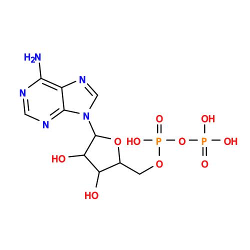 Mass: 427.201 Da / Num. of mol.: 2 / Source method: obtained synthetically / Formula: C10H15N5O10P2 / Feature type: SUBJECT OF INVESTIGATION / Comment: ADP, energy-carrying molecule*YM
 Mass: 24.305 Da / Num. of mol.: 2 / Source method: obtained synthetically / Formula: Mg
 Mass: 40.078 Da / Num. of mol.: 5 / Source method: obtained synthetically / Formula: Ca
 Mass: 18.015 Da / Num. of mol.: 29 / Source method: isolated from a natural source / Formula: H2O
 Sample preparation
 Processing