- PDB-8w1d: CRYSTAL STRUCTURE OF DPS-LIKE PROTEIN PA4880 FROM PSEUDOMONAS AER... -
+
データを開く
IDまたはキーワード:
読み込み中...
-
基本情報
登録情報
データベース: PDB / ID: 8w1d
タイトル
CRYSTAL STRUCTURE OF DPS-LIKE PROTEIN PA4880 FROM PSEUDOMONAS AERUGINOSA (DIMERIC FORM)
要素
DPS-LIKE PROTEIN
キーワード
METAL BINDING PROTEIN / PA4880 / DPS protein / metal binding / DNA cleavage
機能・相同性
機能・相同性情報
ferroxidase / ferroxidase activity / ferric iron binding / iron ion transport / intracellular iron ion homeostasis / iron ion binding / heme binding / cytosol 類似検索 - 分子機能
 ムービー
ムービー コントローラー


 データを開く
 基本情報
基本情報 要素
要素 キーワード
キーワード 機能・相同性情報
 Pseudomonas aeruginosa PAO1 (緑膿菌)
 X線回折 /
  データ登録者
 米国, 1件
  引用
引用 構造の表示
構造の表示 ダウンロードとリンク
 その他のダウンロード

 PDBj
PDBj


 集合体
集合体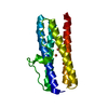


 分子量: 55.845 Da / 分子数: 2 / 由来タイプ: 合成 / 式: Fe / タイプ: SUBJECT OF INVESTIGATION
 分子量: 18.015 Da / 分子数: 147 / 由来タイプ: 天然 / 式: H2O
 試料調製
試料調製 解析
解析