 Movie
Movie Controller


+ Open data

- Basic information
| Entry | Database: PDB / ID: 8w0n | ||||||
|---|---|---|---|---|---|---|---|
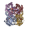
| Title | IRED crystal structure | ||||||
 Components Components | IRED | ||||||
 Keywords Keywords | OXIDOREDUCTASE / protein engineering / imine reductase / scalable manufacturing | ||||||
| Function / homology | NICOTINAMIDE-ADENINE-DINUCLEOTIDE / DI(HYDROXYETHYL)ETHER / PHOSPHATE ION Function and homology information Function and homology information | ||||||
| Biological species |  | ||||||
| Method |  X-RAY DIFFRACTION / SYNCHROTRON / MOLECULAR REPLACEMENT / Resolution: 1.37 Å X-RAY DIFFRACTION / SYNCHROTRON / MOLECULAR REPLACEMENT / Resolution: 1.37 Å | ||||||
 Authors Authors | Peat, T.S. / Newman, J. | ||||||
| Funding support | 1items
| ||||||
 Citation Citation | Journal: Acs Catalysis / Year: 2024 Title: Enhancing the Imine Reductase Activity of a Promiscuous Glucose Dehydrogenase for Scalable Manufacturing of a Chiral Neprilysin Inhibitor Precursor Authors: Yi, X. / Kleinbeck, F. / Ching, C. / Boghospor, L. / Gomes, S. / Alvizo, O. / Allmendinger, T. / Fell, J. / Subramanian, N. / Li, M. / Garcia, R. / Riggins, J. / Entwistle, D. / Richter, Y. ...Authors: Yi, X. / Kleinbeck, F. / Ching, C. / Boghospor, L. / Gomes, S. / Alvizo, O. / Allmendinger, T. / Fell, J. / Subramanian, N. / Li, M. / Garcia, R. / Riggins, J. / Entwistle, D. / Richter, Y. / Gschwend, D. / Lauener, L. / Peat, T.S. / Lebhar, H. / Schlama, T. / Ruch, T. | ||||||
| History |
|
- Structure visualization
| Structure viewer | Molecule: MolmilJmol/JSmol |
|---|
- Downloads & links
-Download
| PDBx/mmCIF format | 8w0n.cif.gz | 306.5 KB | Display | PDBx/mmCIF format |
|---|---|---|---|---|
| PDB format | pdb8w0n.ent.gz | 190 KB | Display | PDB format |
| PDBx/mmJSON format | 8w0n.json.gz | Tree view | PDBx/mmJSON format | |
| Others |  Other downloads Other downloads |
-Validation report
| Arichive directory | https://data.pdbj.org/pub/pdb/validation_reports/w0/8w0nftp://data.pdbj.org/pub/pdb/validation_reports/w0/8w0n | HTTPS FTP |
|---|
-Related structure data
-Links
 PDBj
PDBj- Assembly
| Deposited unit | 
| |||||||||||||||
|---|---|---|---|---|---|---|---|---|---|---|---|---|---|---|---|---|
| 1 | 
| |||||||||||||||
| Unit cell |
| |||||||||||||||
| Components on special symmetry positions |
| |||||||||||||||
| Noncrystallographic symmetry (NCS) | NCS domain:
NCS domain segments: Component-ID: 1 / Ens-ID: 1 / Beg auth comp-ID: MET / Beg label comp-ID: MET / End auth comp-ID: PRO / End label comp-ID: PRO / Auth asym-ID: A / Label asym-ID: A / Auth seq-ID: 1 - 258 / Label seq-ID: 1 - 258
NCS ensembles : (Details: Local NCS retraints between domains: 1 2) |
-Components
-Protein , 1 types, 2 molecules AB
| #1: Protein | Mass: 28068.994 Da / Num. of mol.: 2 Source method: isolated from a genetically manipulated source Source: (gene. exp.) |
|---|
-Non-polymers , 5 types, 467 molecules 


| #2: Chemical |  Mass: 663.425 Da / Num. of mol.: 2 / Source method: obtained synthetically / Formula: C21H27N7O14P2 / Comment: NAD*YM Mass: 663.425 Da / Num. of mol.: 2 / Source method: obtained synthetically / Formula: C21H27N7O14P2 / Comment: NAD*YM#3: Chemical | ChemComp-DMS / |  Mass: 78.133 Da / Num. of mol.: 1 / Source method: obtained synthetically / Formula: C2H6OS / Comment: DMSO, precipitant*YM Mass: 78.133 Da / Num. of mol.: 1 / Source method: obtained synthetically / Formula: C2H6OS / Comment: DMSO, precipitant*YM#4: Chemical | ChemComp-PEG / |  Mass: 106.120 Da / Num. of mol.: 1 / Source method: obtained synthetically / Formula: C4H10O3 Mass: 106.120 Da / Num. of mol.: 1 / Source method: obtained synthetically / Formula: C4H10O3#5: Chemical | ChemComp-PO4 / |  Mass: 94.971 Da / Num. of mol.: 1 / Source method: obtained synthetically / Formula: PO4 Mass: 94.971 Da / Num. of mol.: 1 / Source method: obtained synthetically / Formula: PO4#6: Water | ChemComp-HOH / | Mass: 18.015 Da / Num. of mol.: 462 / Source method: isolated from a natural source / Formula: H2O |
|---|
-Details
| Has ligand of interest | N |
|---|
-Experimental details
-Experiment
| Experiment | Method: X-RAY DIFFRACTION / Number of used crystals: 1 |
|---|
- Sample preparation
| Crystal | Density Matthews: 2.21 Å3/Da / Density % sol: 44.4 % |
|---|---|
| Crystal grow | Temperature: 295 K / Method: vapor diffusion, sitting drop / pH: 5.1 Details: Crystals were found in 20% PEG3350 with 200 mM sodium dihydrogen phosphate when set up in sitting drop plates with 200 nL reservoir and 200 nL of 12 mg/mL protein in SD-2 plates at room temperature. |
-Data collection
| Diffraction | Mean temperature: 100 K / Serial crystal experiment: N |
|---|---|
| Diffraction source | Source: SYNCHROTRON / Site: Australian Synchrotron  / Beamline: MX2 / Wavelength: 0.9537 Å / Beamline: MX2 / Wavelength: 0.9537 Å |
| Detector | Type: DECTRIS EIGER X 16M / Detector: PIXEL / Date: Mar 18, 2023 |
| Radiation | Protocol: SINGLE WAVELENGTH / Monochromatic (M) / Laue (L): M / Scattering type: x-ray |
| Radiation wavelength | Wavelength: 0.9537 Å / Relative weight: 1 |
| Reflection | Resolution: 1.37→48.5 Å / Num. obs: 102543 / % possible obs: 99.6 % / Redundancy: 20.3 % / CC1/2: 0.998 / Rmerge(I) obs: 0.1 / Rpim(I) all: 0.023 / Net I/σ(I): 17.6 |
| Reflection shell | Resolution: 1.37→1.39 Å / Redundancy: 18 % / Rmerge(I) obs: 0.985 / Mean I/σ(I) obs: 3.9 / Num. unique obs: 4726 / CC1/2: 0.914 / Rpim(I) all: 0.233 / % possible all: 92.7 |
- Processing
| Software |
| |||||||||||||||||||||||||||||||||||||||||||||||||||||||||||||||||||||||||||||||||||||||||||||||||||||||||||||||||||||||||||||||||||||||||||||||||||||||||||||||||||||
|---|---|---|---|---|---|---|---|---|---|---|---|---|---|---|---|---|---|---|---|---|---|---|---|---|---|---|---|---|---|---|---|---|---|---|---|---|---|---|---|---|---|---|---|---|---|---|---|---|---|---|---|---|---|---|---|---|---|---|---|---|---|---|---|---|---|---|---|---|---|---|---|---|---|---|---|---|---|---|---|---|---|---|---|---|---|---|---|---|---|---|---|---|---|---|---|---|---|---|---|---|---|---|---|---|---|---|---|---|---|---|---|---|---|---|---|---|---|---|---|---|---|---|---|---|---|---|---|---|---|---|---|---|---|---|---|---|---|---|---|---|---|---|---|---|---|---|---|---|---|---|---|---|---|---|---|---|---|---|---|---|---|---|---|---|---|---|
| Refinement | Method to determine structure: MOLECULAR REPLACEMENT / Resolution: 1.37→48.5 Å / Cor.coef. Fo:Fc: 0.982 / Cor.coef. Fo:Fc free: 0.976 / SU B: 1.327 / SU ML: 0.025 / Cross valid method: FREE R-VALUE / ESU R: 0.048 / ESU R Free: 0.044 Details: Hydrogens have been added in their riding positions
| |||||||||||||||||||||||||||||||||||||||||||||||||||||||||||||||||||||||||||||||||||||||||||||||||||||||||||||||||||||||||||||||||||||||||||||||||||||||||||||||||||||
| Solvent computation | Ion probe radii: 0.8 Å / Shrinkage radii: 0.8 Å / VDW probe radii: 1.2 Å / Solvent model: MASK BULK SOLVENT | |||||||||||||||||||||||||||||||||||||||||||||||||||||||||||||||||||||||||||||||||||||||||||||||||||||||||||||||||||||||||||||||||||||||||||||||||||||||||||||||||||||
| Displacement parameters | Biso mean: 16.548 Å2
| |||||||||||||||||||||||||||||||||||||||||||||||||||||||||||||||||||||||||||||||||||||||||||||||||||||||||||||||||||||||||||||||||||||||||||||||||||||||||||||||||||||
| Refinement step | Cycle: LAST / Resolution: 1.37→48.5 Å
| |||||||||||||||||||||||||||||||||||||||||||||||||||||||||||||||||||||||||||||||||||||||||||||||||||||||||||||||||||||||||||||||||||||||||||||||||||||||||||||||||||||
| Refine LS restraints |
| |||||||||||||||||||||||||||||||||||||||||||||||||||||||||||||||||||||||||||||||||||||||||||||||||||||||||||||||||||||||||||||||||||||||||||||||||||||||||||||||||||||
| Refine LS restraints NCS |
| |||||||||||||||||||||||||||||||||||||||||||||||||||||||||||||||||||||||||||||||||||||||||||||||||||||||||||||||||||||||||||||||||||||||||||||||||||||||||||||||||||||
| LS refinement shell |
|
