 Movie
Movie Controller

[English] 日本語
 Yorodumi
- PDB-8vzx: Q108K:K40L:T53A:R58F mutant of hCRBPII bound to synthetic fluorop... -
+ Open data

- Basic information
| Entry | Database: PDB / ID: 8vzx | ||||||
|---|---|---|---|---|---|---|---|
| Title | Q108K:K40L:T53A:R58F mutant of hCRBPII bound to synthetic fluorophore TD-1V | ||||||
 Components Components | Retinol-binding protein 2 | ||||||
 Keywords Keywords | RETINOL BINDING PROTEIN / human cellular retinol binding protein II / hCRBPII / TD-1V / engineered protein / fluorescent protein | ||||||
| Function / homology |  Function and homology information Function and homology informationvitamin A metabolic process / retinoid binding / retinal binding / retinol binding / epidermis development / fatty acid transport / Retinoid metabolism and transport / fatty acid binding / nucleus / cytosol Similarity search - Function | ||||||
| Biological species |  Homo sapiens (human) Homo sapiens (human) | ||||||
| Method |  X-RAY DIFFRACTION / SYNCHROTRON / MOLECULAR REPLACEMENT / Resolution: 1.47 Å X-RAY DIFFRACTION / SYNCHROTRON / MOLECULAR REPLACEMENT / Resolution: 1.47 Å | ||||||
 Authors Authors | Nossoni, Z. / Bingham, C.R. / Geiger, J.H. | ||||||
| Funding support |  United States, 1items United States, 1items
| ||||||
 Citation Citation | Journal: Acs Chem.Biol. / Year: 2024 Title: Regulation of Absorption and Emission in a Protein/Fluorophore Complex. Authors: Santos, E.M. / Chandra, I. / Assar, Z. / Sheng, W. / Ghanbarpour, A. / Bingham, C. / Vasileiou, C. / Geiger, J.H. / Borhan, B. | ||||||
| History |
|
- Structure visualization
| Structure viewer | Molecule: MolmilJmol/JSmol |
|---|
- Downloads & links
-Download
| PDBx/mmCIF format | 8vzx.cif.gz | 256 KB | Display | PDBx/mmCIF format |
|---|---|---|---|---|
| PDB format | pdb8vzx.ent.gz | Display | PDB format | |
| PDBx/mmJSON format | 8vzx.json.gz | Tree view | PDBx/mmJSON format | |
| Others |  Other downloads Other downloads |
-Validation report
| Arichive directory | https://data.pdbj.org/pub/pdb/validation_reports/vz/8vzxftp://data.pdbj.org/pub/pdb/validation_reports/vz/8vzx | HTTPS FTP |
|---|
-Related structure data
| Related structure data | 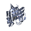 8vzyC  8vzzC  8w00C 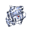 8w02C  4qynS S: Starting model for refinement C: citing same article ( |
|---|---|
| Similar structure data |
-Links
 PDBj
PDBj


- Assembly
| Deposited unit | 
| ||||||||||||
|---|---|---|---|---|---|---|---|---|---|---|---|---|---|
| 1 | 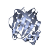
| ||||||||||||
| 2 | 
| ||||||||||||
| 3 | 
| ||||||||||||
| 4 | 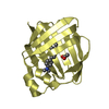
| ||||||||||||
| Unit cell |
|
-Components
| #1: Protein | Mass: 15542.435 Da / Num. of mol.: 4 / Mutation: Q108K:K40L:T53A:R58F Source method: isolated from a genetically manipulated source Source: (gene. exp.) Homo sapiens (human) / Gene: RBP2, CRBP2 / Production host:  #2: Chemical | ChemComp-ACT /   Mass: 59.044 Da / Num. of mol.: 4 / Source method: obtained synthetically / Formula: C2H3O2 Mass: 59.044 Da / Num. of mol.: 4 / Source method: obtained synthetically / Formula: C2H3O2#3: Chemical | Mass: 271.377 Da / Num. of mol.: 2 / Source method: obtained synthetically / Formula: C16H17NOS / Feature type: SUBJECT OF INVESTIGATION #4: Water | ChemComp-HOH / |  Mass: 18.015 Da / Num. of mol.: 402 / Source method: isolated from a natural source / Formula: H2O Mass: 18.015 Da / Num. of mol.: 402 / Source method: isolated from a natural source / Formula: H2OHas ligand of interest | Y | Has protein modification | Y | |
|---|
-Experimental details
-Experiment
| Experiment | Method: X-RAY DIFFRACTION / Number of used crystals: 1 |
|---|
- Sample preparation
| Crystal | Density Matthews: 2.03 Å3/Da / Density % sol: 39.26 % |
|---|---|
| Crystal grow | Temperature: 295 K / Method: vapor diffusion, hanging drop / Details: ammonium acetate, sodium acetate, PEG4000 / PH range: 4.0-4.8 / Temp details: room temperature |
-Data collection
| Diffraction | Mean temperature: 100 K / Serial crystal experiment: N |
|---|---|
| Diffraction source | Source: SYNCHROTRON / Site: APS / Beamline: 21-ID-F / Wavelength: 0.97872 Å |
| Detector | Type: DECTRIS EIGER X 9M / Detector: PIXEL / Date: Sep 17, 2015 |
| Radiation | Protocol: SINGLE WAVELENGTH / Monochromatic (M) / Laue (L): M / Scattering type: x-ray |
| Radiation wavelength | Wavelength: 0.97872 Å / Relative weight: 1 |
| Reflection | Resolution: 1.47→27.4 Å / Num. obs: 82805 / % possible obs: 95.71 % / Redundancy: 2.9 % / CC1/2: 0.896 / Net I/σ(I): 32.9 |
| Reflection shell | Resolution: 1.47→1.51 Å / Num. unique obs: 16538 / CC1/2: 0.912 |
- Processing
| Software |
| ||||||||||||||||||||||||
|---|---|---|---|---|---|---|---|---|---|---|---|---|---|---|---|---|---|---|---|---|---|---|---|---|---|
| Refinement | Method to determine structure: MOLECULAR REPLACEMENT Starting model: PDB entry 4QYN Resolution: 1.47→27.4 Å / Cross valid method: FREE R-VALUE Stereochemistry target values: GeoStd + Monomer Library + CDL v1.2
| ||||||||||||||||||||||||
| Displacement parameters | Biso mean: 26.56 Å2 | ||||||||||||||||||||||||
| Refinement step | Cycle: LAST / Resolution: 1.47→27.4 Å
| ||||||||||||||||||||||||
| Refine LS restraints |
|
