 Movie
Movie Controller


+ Open data

- Basic information
| Entry | Database: PDB / ID: 8vrv | ||||||
|---|---|---|---|---|---|---|---|
| Title | GII.4c H2-tri HBGA norovirus protruding domain | ||||||
 Components Components | Capsid protein | ||||||
 Keywords Keywords | VIRAL PROTEIN / norovirus | ||||||
| Function / homology |  Function and homology information Function and homology information | ||||||
| Biological species |  Norovirus GII.4 Norovirus GII.4 | ||||||
| Method |  X-RAY DIFFRACTION / SYNCHROTRON / MOLECULAR REPLACEMENT / Resolution: 1.61 Å X-RAY DIFFRACTION / SYNCHROTRON / MOLECULAR REPLACEMENT / Resolution: 1.61 Å | ||||||
 Authors Authors | Kher, G. / Prewitt, A. / Pancera, M. / Hansman, G. | ||||||
| Funding support |  Germany, 1items Germany, 1items
| ||||||
 Citation Citation | Journal: J.Virol. / Year: 2024 Title: Development of a broad-spectrum therapeutic Fc-nanobody for human noroviruses. Authors: Hansman, G.S. / Kher, G. / Svirina, A.D. / Tame, J.R.H. / Hartley-Tassell, L. / Irie, H. / Haselhorst, T. / von Itzstein, M. / Rudd, P.A. / Pancera, M. | ||||||
| History |
|
- Structure visualization
| Structure viewer | Molecule: MolmilJmol/JSmol |
|---|
- Downloads & links
-Download
| PDBx/mmCIF format | 8vrv.cif.gz | 242.7 KB | Display | PDBx/mmCIF format |
|---|---|---|---|---|
| PDB format | pdb8vrv.ent.gz | Display | PDB format | |
| PDBx/mmJSON format | 8vrv.json.gz | Tree view | PDBx/mmJSON format | |
| Others |  Other downloads Other downloads |
-Validation report
| Arichive directory | https://data.pdbj.org/pub/pdb/validation_reports/vr/8vrvftp://data.pdbj.org/pub/pdb/validation_reports/vr/8vrv | HTTPS FTP |
|---|
-Related structure data
| Related structure data | 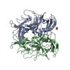 8v95C 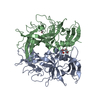 8v96C 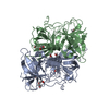 8v97C 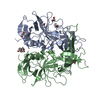 8v98C 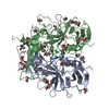 8v99C 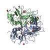 8v9aC 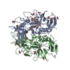 8vruC C: citing same article ( |
|---|---|
| Similar structure data |
-Links
 PDBj
PDBj
- Assembly
| Deposited unit | 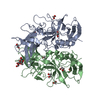
| ||||||||
|---|---|---|---|---|---|---|---|---|---|
| 1 |
| ||||||||
| Unit cell |
|
-Components
| #1: Protein | Mass: 33667.480 Da / Num. of mol.: 2 Source method: isolated from a genetically manipulated source Source: (gene. exp.) Norovirus GII.4 / Production host:  #2: Polysaccharide | alpha-L-fucopyranose-(1-2)-beta-D-galactopyranose | #3: Chemical | ChemComp-EDO /   Mass: 62.068 Da / Num. of mol.: 6 / Source method: obtained synthetically / Formula: C2H6O2 Mass: 62.068 Da / Num. of mol.: 6 / Source method: obtained synthetically / Formula: C2H6O2#4: Chemical |   Mass: 46.025 Da / Num. of mol.: 3 / Source method: obtained synthetically / Formula: CH2O2 Mass: 46.025 Da / Num. of mol.: 3 / Source method: obtained synthetically / Formula: CH2O2#5: Water | ChemComp-HOH / |  Mass: 18.015 Da / Num. of mol.: 669 / Source method: isolated from a natural source / Formula: H2O Mass: 18.015 Da / Num. of mol.: 669 / Source method: isolated from a natural source / Formula: H2OHas ligand of interest | Y | Has protein modification | N | |
|---|
-Experimental details
-Experiment
| Experiment | Method: X-RAY DIFFRACTION / Number of used crystals: 1 |
|---|
- Sample preparation
| Crystal | Density Matthews: 2.35 Å3/Da / Density % sol: 47.55 % |
|---|---|
| Crystal grow | Temperature: 291.15 K / Method: vapor diffusion, hanging drop Details: SG1 1-24 0.2 M Magnesium formate dihydrate 20% w/v PEG 3350 |
-Data collection
| Diffraction | Mean temperature: 100 K / Serial crystal experiment: N |
|---|---|
| Diffraction source | Source: SYNCHROTRON / Site: Australian Synchrotron  / Beamline: MX1 / Wavelength: 0.95373 Å / Beamline: MX1 / Wavelength: 0.95373 Å |
| Detector | Type: DECTRIS EIGER2 X 9M / Detector: PIXEL / Date: Apr 14, 2023 |
| Radiation | Protocol: SINGLE WAVELENGTH / Monochromatic (M) / Laue (L): M / Scattering type: x-ray |
| Radiation wavelength | Wavelength: 0.95373 Å / Relative weight: 1 |
| Reflection | Resolution: 1.61→49.26 Å / Num. obs: 82708 / % possible obs: 99.9 % / Redundancy: 6.8 % / CC1/2: 0.993 / Rmerge(I) obs: 0.077 / Rpim(I) all: 0.032 / Rrim(I) all: 0.083 / Χ2: 0.56 / Net I/σ(I): 11.7 / Num. measured all: 562066 |
| Reflection shell | Resolution: 1.61→1.64 Å / % possible obs: 98.5 % / Redundancy: 6.6 % / Rmerge(I) obs: 0.797 / Num. measured all: 26494 / Num. unique obs: 3989 / CC1/2: 0.769 / Rpim(I) all: 0.326 / Rrim(I) all: 0.862 / Χ2: 0.51 / Net I/σ(I) obs: 1.7 |
- Processing
| Software |
| |||||||||||||||||||||||||||||||||||||||||||||||||||||||||||||||||||||||||||||||||||||||||||||||||||||||||||||||||||||||||||||||||||||||||||||||||||||||||||||||||||||||||||||||||||||||||||||||||||||||||||||||||||||||||
|---|---|---|---|---|---|---|---|---|---|---|---|---|---|---|---|---|---|---|---|---|---|---|---|---|---|---|---|---|---|---|---|---|---|---|---|---|---|---|---|---|---|---|---|---|---|---|---|---|---|---|---|---|---|---|---|---|---|---|---|---|---|---|---|---|---|---|---|---|---|---|---|---|---|---|---|---|---|---|---|---|---|---|---|---|---|---|---|---|---|---|---|---|---|---|---|---|---|---|---|---|---|---|---|---|---|---|---|---|---|---|---|---|---|---|---|---|---|---|---|---|---|---|---|---|---|---|---|---|---|---|---|---|---|---|---|---|---|---|---|---|---|---|---|---|---|---|---|---|---|---|---|---|---|---|---|---|---|---|---|---|---|---|---|---|---|---|---|---|---|---|---|---|---|---|---|---|---|---|---|---|---|---|---|---|---|---|---|---|---|---|---|---|---|---|---|---|---|---|---|---|---|---|---|---|---|---|---|---|---|---|---|---|---|---|---|---|---|---|
| Refinement | Method to determine structure: MOLECULAR REPLACEMENT / Resolution: 1.61→45.67 Å / SU ML: 0.14 / Cross valid method: FREE R-VALUE / σ(F): 1.35 / Phase error: 16.9 / Stereochemistry target values: ML
| |||||||||||||||||||||||||||||||||||||||||||||||||||||||||||||||||||||||||||||||||||||||||||||||||||||||||||||||||||||||||||||||||||||||||||||||||||||||||||||||||||||||||||||||||||||||||||||||||||||||||||||||||||||||||
| Solvent computation | Shrinkage radii: 0.9 Å / VDW probe radii: 1.11 Å / Solvent model: FLAT BULK SOLVENT MODEL | |||||||||||||||||||||||||||||||||||||||||||||||||||||||||||||||||||||||||||||||||||||||||||||||||||||||||||||||||||||||||||||||||||||||||||||||||||||||||||||||||||||||||||||||||||||||||||||||||||||||||||||||||||||||||
| Refinement step | Cycle: LAST / Resolution: 1.61→45.67 Å
| |||||||||||||||||||||||||||||||||||||||||||||||||||||||||||||||||||||||||||||||||||||||||||||||||||||||||||||||||||||||||||||||||||||||||||||||||||||||||||||||||||||||||||||||||||||||||||||||||||||||||||||||||||||||||
| Refine LS restraints |
| |||||||||||||||||||||||||||||||||||||||||||||||||||||||||||||||||||||||||||||||||||||||||||||||||||||||||||||||||||||||||||||||||||||||||||||||||||||||||||||||||||||||||||||||||||||||||||||||||||||||||||||||||||||||||
| LS refinement shell |
|
