 Movie
Movie Controller

[English] 日本語
 Yorodumi
- PDB-8vr9: Structure of a synthetic antibody in complex with a class I MHC p... -
+ Open data

- Basic information
| Entry | Database: PDB / ID: 8vr9 | ||||||
|---|---|---|---|---|---|---|---|
| Title | Structure of a synthetic antibody in complex with a class I MHC presenting a hapten-peptide conjugate | ||||||
 Components Components |
| ||||||
 Keywords Keywords | IMMUNE SYSTEM / Complex / Synthetic antibody / MHC class I / covalently modified peptide | ||||||
| Function / homology |  Function and homology information Function and homology informationpositive regulation of memory T cell activation / T cell mediated cytotoxicity directed against tumor cell target / positive regulation of CD8-positive, alpha-beta T cell activation / CD8-positive, alpha-beta T cell activation / positive regulation of CD8-positive, alpha-beta T cell proliferation / response to mineralocorticoid / GMP binding / forebrain astrocyte development / antigen processing and presentation of endogenous peptide antigen via MHC class I via ER pathway, TAP-dependent / TAP complex binding ...positive regulation of memory T cell activation / T cell mediated cytotoxicity directed against tumor cell target / positive regulation of CD8-positive, alpha-beta T cell activation / CD8-positive, alpha-beta T cell activation / positive regulation of CD8-positive, alpha-beta T cell proliferation / response to mineralocorticoid / GMP binding / forebrain astrocyte development / antigen processing and presentation of endogenous peptide antigen via MHC class I via ER pathway, TAP-dependent / TAP complex binding / antigen processing and presentation of exogenous peptide antigen via MHC class I / LRR domain binding / Golgi medial cisterna / regulation of synaptic transmission, GABAergic / negative regulation of epithelial cell differentiation / response to isolation stress / response to gravity / epithelial tube branching involved in lung morphogenesis / CD8 receptor binding / type I pneumocyte differentiation / protection from natural killer cell mediated cytotoxicity / Rac protein signal transduction / beta-2-microglobulin binding / endoplasmic reticulum exit site / myoblast proliferation / Signaling by RAS GAP mutants / Signaling by RAS GTPase mutants / Activation of RAS in B cells / TAP binding / RAS signaling downstream of NF1 loss-of-function variants / RUNX3 regulates p14-ARF / skeletal muscle cell differentiation / positive regulation of glial cell proliferation / SOS-mediated signalling / detection of bacterium / Activated NTRK3 signals through RAS / Activated NTRK2 signals through RAS / antigen processing and presentation of endogenous peptide antigen via MHC class Ib / antigen processing and presentation of endogenous peptide antigen via MHC class I via ER pathway, TAP-independent / cardiac muscle cell proliferation / SHC1 events in ERBB4 signaling / Signalling to RAS / SHC-related events triggered by IGF1R / Activated NTRK2 signals through FRS2 and FRS3 / Estrogen-stimulated signaling through PRKCZ / positive regulation of Rac protein signal transduction / SHC-mediated cascade:FGFR3 / glial cell proliferation / MET activates RAS signaling / SHC-mediated cascade:FGFR2 / SHC-mediated cascade:FGFR4 / Signaling by PDGFRA transmembrane, juxtamembrane and kinase domain mutants / Signaling by PDGFRA extracellular domain mutants / PTK6 Regulates RHO GTPases, RAS GTPase and MAP kinases / Erythropoietin activates RAS / SHC-mediated cascade:FGFR1 / Signaling by FGFR4 in disease / T cell receptor binding / Signaling by CSF3 (G-CSF) / FRS-mediated FGFR3 signaling / Signaling by FLT3 ITD and TKD mutants / FRS-mediated FGFR2 signaling / FRS-mediated FGFR4 signaling / p38MAPK events / striated muscle cell differentiation / FRS-mediated FGFR1 signaling / Signaling by FGFR3 in disease / Tie2 Signaling / protein-membrane adaptor activity / Signaling by FGFR2 in disease / Signaling by FLT3 fusion proteins / GRB2 events in EGFR signaling / SHC1 events in EGFR signaling / FLT3 Signaling / Signaling by FGFR1 in disease / EGFR Transactivation by Gastrin / NCAM signaling for neurite out-growth / CD209 (DC-SIGN) signaling / Downstream signal transduction / GRB2 events in ERBB2 signaling / homeostasis of number of cells within a tissue / Insulin receptor signalling cascade / SHC1 events in ERBB2 signaling / early endosome lumen / Nef mediated downregulation of MHC class I complex cell surface expression / DAP12 interactions / Constitutive Signaling by Overexpressed ERBB2 / Signaling by phosphorylated juxtamembrane, extracellular and kinase domain KIT mutants / Ras activation upon Ca2+ influx through NMDA receptor / response to glucocorticoid / Endosomal/Vacuolar pathway / T cell mediated cytotoxicity / VEGFR2 mediated cell proliferation / small monomeric GTPase / Antigen Presentation: Folding, assembly and peptide loading of class I MHC / lumenal side of endoplasmic reticulum membrane / regulation of iron ion transport / FCERI mediated MAPK activation / cellular response to iron(III) ion / negative regulation of iron ion transport Similarity search - Function | ||||||
| Biological species |  Homo sapiens (human) Homo sapiens (human) | ||||||
| Method | ELECTRON MICROSCOPY / single particle reconstruction / cryo EM / Resolution: 3.06 Å | ||||||
 Authors Authors | Maso, L. / Bang, I. / Koide, S. | ||||||
| Funding support |  United States, 1items United States, 1items
| ||||||
 Citation Citation | Journal: Proc Natl Acad Sci U S A / Year: 2024 Title: Molecular basis for antibody recognition of multiple drug-peptide/MHC complexes. Authors: Lorenzo Maso / Epsa Rajak / Injin Bang / Akiko Koide / Takamitsu Hattori / Benjamin G Neel / Shohei Koide / Abstract: The HapImmune platform exploits covalent inhibitors as haptens for creating major histocompatibility complex (MHC)-presented tumor-specific neoantigens by design, combining targeted therapies with ...The HapImmune platform exploits covalent inhibitors as haptens for creating major histocompatibility complex (MHC)-presented tumor-specific neoantigens by design, combining targeted therapies with immunotherapy for the treatment of drug-resistant cancers. A HapImmune antibody, R023, recognizes multiple sotorasib-conjugated KRAS(G12C) peptides presented by different human leukocyte antigens (HLAs). This high specificity to sotorasib, coupled with broad HLA-binding capability, enables such antibodies, when reformatted as T cell engagers, to potently and selectively kill sotorasib-resistant KRAS(G12C) cancer cells expressing different HLAs upon sotorasib treatment. The loosening of HLA restriction could increase the patient population that can benefit from this therapeutic approach. To understand the molecular basis for its unconventional binding capability, we used single-particle cryogenic electron microscopy to determine the structures of R023 bound to multiple sotorasib-peptide conjugates presented by different HLAs. R023 forms a pocket for sotorasib between the V and V domains, binds HLAs in an unconventional, angled way, with V making most contacts with them, and makes few contacts with the peptide moieties. This binding mode enables the antibody to accommodate different hapten-peptide conjugates and to adjust its conformation to different HLAs presenting hapten-peptides. Deep mutational scanning validated the structures and revealed distinct levels of mutation tolerance by sotorasib- and HLA-binding residues. Together, our structural information and sequence landscape analysis reveal key features for achieving MHC-restricted recognition of multiple hapten-peptide antigens, which will inform the development of next-generation therapeutic antibodies. | ||||||
| History |
|
- Structure visualization
| Structure viewer | Molecule: MolmilJmol/JSmol |
|---|
- Downloads & links
-Download
| PDBx/mmCIF format | 8vr9.cif.gz | 125.5 KB | Display | PDBx/mmCIF format |
|---|---|---|---|---|
| PDB format | pdb8vr9.ent.gz | 92.6 KB | Display | PDB format |
| PDBx/mmJSON format | 8vr9.json.gz | Tree view | PDBx/mmJSON format | |
| Others |  Other downloads Other downloads |
-Validation report
| Arichive directory | https://data.pdbj.org/pub/pdb/validation_reports/vr/8vr9ftp://data.pdbj.org/pub/pdb/validation_reports/vr/8vr9 | HTTPS FTP |
|---|
-Related structure data
| Related structure data |  43478MC 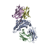 8vraC 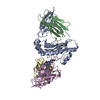 8vrbC M: map data used to model this data C: citing same article ( |
|---|---|
| Similar structure data |
-Links
 PDBj
PDBj


- Assembly
| Deposited unit | 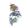
|
|---|---|
| 1 |
|
-Components
-Protein , 2 types, 2 molecules AB
| #1: Protein | Mass: 32047.309 Da / Num. of mol.: 1 / Fragment: extracellular domain Source method: isolated from a genetically manipulated source Source: (gene. exp.) Homo sapiens (human) / Gene: HLA-A, HLAA / Plasmid: pET28 / Production host:  |
|---|---|
| #2: Protein | Mass: 11892.291 Da / Num. of mol.: 1 Source method: isolated from a genetically manipulated source Source: (gene. exp.) Homo sapiens (human) / Gene: B2M, CDABP0092, HDCMA22P / Plasmid: pHFT2 / Production host: |
-Antibody , 2 types, 2 molecules DE
| #4: Antibody | Mass: 23518.178 Da / Num. of mol.: 1 Source method: isolated from a genetically manipulated source Source: (gene. exp.) Homo sapiens (human) / Production host: |
|---|---|
| #5: Antibody | Mass: 24034.822 Da / Num. of mol.: 1 Source method: isolated from a genetically manipulated source Source: (gene. exp.) Homo sapiens (human) / Production host: |
-Protein/peptide / Non-polymers , 2 types, 2 molecules C
| #3: Protein/peptide | Mass: 789.963 Da / Num. of mol.: 1 / Fragment: residues 8-16 / Mutation: G12C / Source method: obtained synthetically / Details: Cys12 conjugated to covalent inhibitor sotorasib / Source: (synth.) Homo sapiens (human) / References: UniProt: P01116 |
|---|---|
| #6: Chemical | ChemComp-MOV /  Mass: 562.610 Da / Num. of mol.: 1 / Source method: obtained synthetically / Formula: C30H32F2N6O3 / Feature type: SUBJECT OF INVESTIGATION Mass: 562.610 Da / Num. of mol.: 1 / Source method: obtained synthetically / Formula: C30H32F2N6O3 / Feature type: SUBJECT OF INVESTIGATION |
-Details
| Has ligand of interest | Y |
|---|---|
| Has protein modification | Y |
-Experimental details
-Experiment
| Experiment | Method: ELECTRON MICROSCOPY |
|---|---|
| EM experiment | Aggregation state: PARTICLE / 3D reconstruction method: single particle reconstruction |
- Sample preparation
| Component |
| ||||||||||||||||||||||||||||||
|---|---|---|---|---|---|---|---|---|---|---|---|---|---|---|---|---|---|---|---|---|---|---|---|---|---|---|---|---|---|---|---|
| Molecular weight | Value: 0.0927 MDa / Experimental value: NO | ||||||||||||||||||||||||||||||
| Source (natural) |
| ||||||||||||||||||||||||||||||
| Source (recombinant) |
| ||||||||||||||||||||||||||||||
| Buffer solution | pH: 7.4 | ||||||||||||||||||||||||||||||
| Buffer component |
| ||||||||||||||||||||||||||||||
| Specimen | Conc.: 1.5 mg/ml / Embedding applied: NO / Shadowing applied: NO / Staining applied: NO / Vitrification applied: YES | ||||||||||||||||||||||||||||||
| Specimen support | Grid material: GOLD / Grid mesh size: 300 divisions/in. / Grid type: Quantifoil R0.6/1 | ||||||||||||||||||||||||||||||
| Vitrification | Instrument: FEI VITROBOT MARK IV / Cryogen name: ETHANE / Humidity: 100 % / Chamber temperature: 277 K |
- Electron microscopy imaging
| Experimental equipment |  Model: Titan Krios / Image courtesy: FEI Company |
|---|---|
| Microscopy | Model: FEI TITAN KRIOS |
| Electron gun | Electron source:  FIELD EMISSION GUN / Accelerating voltage: 300 kV / Illumination mode: FLOOD BEAM FIELD EMISSION GUN / Accelerating voltage: 300 kV / Illumination mode: FLOOD BEAM |
| Electron lens | Mode: BRIGHT FIELD / Nominal magnification: 105000 X / Nominal defocus max: 2400 nm / Nominal defocus min: 900 nm / Cs: 2.7 mm |
| Specimen holder | Cryogen: NITROGEN |
| Image recording | Average exposure time: 2 sec. / Electron dose: 57.72 e/Å2 / Film or detector model: GATAN K3 (6k x 4k) / Num. of grids imaged: 1 |
| EM imaging optics | Energyfilter slit width: 20 eV |
- Processing
| EM software |
| ||||||||||||||||
|---|---|---|---|---|---|---|---|---|---|---|---|---|---|---|---|---|---|
| CTF correction | Type: PHASE FLIPPING AND AMPLITUDE CORRECTION | ||||||||||||||||
| 3D reconstruction | Resolution: 3.06 Å / Resolution method: FSC 0.143 CUT-OFF / Num. of particles: 178888 / Symmetry type: POINT |
