National Institutes of Health/National Institute of Environmental Health Sciences (NIH/NIEHS)
United States
Citation
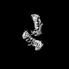
Journal: Nat Commun / Year: 2025 Title: A higher order PUF complex is central to regulation of C. elegans germline stem cells. Authors: Chen Qiu / Sarah L Crittenden / Brian H Carrick / Lucas B Dillard / Stephany J Costa Dos Santos / Venkata P Dandey / Robert C Dutcher / Elizabeth G Viverette / Robert N Wine / Jennifer ...Authors: Chen Qiu / Sarah L Crittenden / Brian H Carrick / Lucas B Dillard / Stephany J Costa Dos Santos / Venkata P Dandey / Robert C Dutcher / Elizabeth G Viverette / Robert N Wine / Jennifer Woodworth / Zachary T Campbell / Marvin Wickens / Mario J Borgnia / Judith Kimble / Traci M Tanaka Hall / Abstract: PUF RNA-binding proteins are broadly conserved stem cell regulators. Nematode PUF proteins maintain germline stem cells (GSCs) and, with key partner proteins, repress differentiation mRNAs, including ...PUF RNA-binding proteins are broadly conserved stem cell regulators. Nematode PUF proteins maintain germline stem cells (GSCs) and, with key partner proteins, repress differentiation mRNAs, including gld-1. Here we report that PUF protein FBF-2 and its partner LST-1 form a ternary complex that represses gld-1 via a pair of adjacent FBF binding elements (FBEs) in its 3'UTR. One LST-1 molecule links two FBF-2 molecules via motifs in the LST-1 intrinsically-disordered region; the gld-1 FBE pair includes a well-established 'canonical' FBE and a newly-identified noncanonical FBE. Remarkably, this FBE pair drives both full RNA repression in GSCs and full RNA activation upon differentiation. Discoveries of the LST-1-FBF-2 ternary complex, the gld-1 adjacent FBEs, and their in vivo significance predict an expanded regulatory repertoire of different assemblies of PUF-partner-RNA higher order complexes in nematode GSCs. This also suggests analogous PUF controls may await discovery in other biological contexts and organisms.
In the structure databanks used in Yorodumi, some data are registered as the other names, "COVID-19 virus" and "2019-nCoV". Here are the details of the virus and the list of structure data.
Jan 31, 2019. EMDB accession codes are about to change! (news from PDBe EMDB page)
EMDB accession codes are about to change! (news from PDBe EMDB page)
The allocation of 4 digits for EMDB accession codes will soon come to an end. Whilst these codes will remain in use, new EMDB accession codes will include an additional digit and will expand incrementally as the available range of codes is exhausted. The current 4-digit format prefixed with “EMD-” (i.e. EMD-XXXX) will advance to a 5-digit format (i.e. EMD-XXXXX), and so on. It is currently estimated that the 4-digit codes will be depleted around Spring 2019, at which point the 5-digit format will come into force.
The EM Navigator/Yorodumi systems omit the EMD- prefix.
Related info.:Q: What is EMD? / ID/Accession-code notation in Yorodumi/EM Navigator
Yorodumi is a browser for structure data from EMDB, PDB, SASBDB, etc.
This page is also the successor to EM Navigator detail page, and also detail information page/front-end page for Omokage search.
The word "yorodu" (or yorozu) is an old Japanese word meaning "ten thousand". "mi" (miru) is to see.
Related info.:EMDB / PDB / SASBDB / Comparison of 3 databanks / Yorodumi Search / Aug 31, 2016. New EM Navigator & Yorodumi / Yorodumi Papers / Jmol/JSmol / Function and homology information / Changes in new EM Navigator and Yorodumi
 Movie
Movie Controller


 Open data
 Basic information
 Components
 Keywords
 Function and homology information
 X-RAY DIFFRACTION /
  Authors
 United States, 1items
  Citation
 Structure visualization
 Downloads & links
 Other downloads
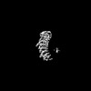
 PDBj
PDBj


 Assembly

 Mass: 18.015 Da / Num. of mol.: 92 / Source method: isolated from a natural source / Formula: H2O
 Sample preparation
 Processing