 Movie
Movie Controller

[English] 日本語
 Yorodumi
- PDB-8vig: EgtB-IV from Geminocystis sp. isolate SKYG4, an ergothioneine-bio... -
+ Open data

- Basic information
| Entry | Database: PDB / ID: 8vig | |||||||||
|---|---|---|---|---|---|---|---|---|---|---|
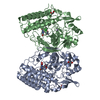
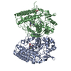
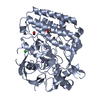
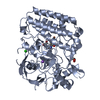
| Title | EgtB-IV from Geminocystis sp. isolate SKYG4, an ergothioneine-biosynthetic type IV sulfoxide synthase in complex with hercynine | |||||||||
 Components Components | Sulfoxide synthase EgtB-IV | |||||||||
 Keywords Keywords | OXIDOREDUCTASE / ergothioneine / sulfoxide / sulfur / non-heme iron | |||||||||
| Function / homology | 5-histidylcysteine sulfoxide synthase / : / Sulfatase-modifying factor enzyme / Sulfatase-modifying factor enzyme 1-like domain / Sulfatase-modifying factor enzyme superfamily / C-type lectin fold / N,N,N-trimethyl-histidine / : / 5-histidylcysteine sulfoxide synthase Function and homology information Function and homology information | |||||||||
| Biological species |  Geminocystis sp. (bacteria) Geminocystis sp. (bacteria) | |||||||||
| Method |  X-RAY DIFFRACTION / SYNCHROTRON / MOLECULAR REPLACEMENT / Resolution: 1.6 Å X-RAY DIFFRACTION / SYNCHROTRON / MOLECULAR REPLACEMENT / Resolution: 1.6 Å | |||||||||
 Authors Authors | Ireland, K.A. / Davis, K.M. | |||||||||
| Funding support |  United States, 2items United States, 2items
| |||||||||
 Citation Citation | Journal: Structure / Year: 2024 Title: Structural insights into the convergent evolution of sulfoxide synthase EgtB-IV, an ergothioneine-biosynthetic homolog of ovothiol synthase OvoA. Authors: Ireland, K.A. / Kayrouz, C.M. / Abbott, M.L. / Seyedsayamdost, M.R. / Davis, K.M. | |||||||||
| History |
|
- Structure visualization
| Structure viewer | Molecule: MolmilJmol/JSmol |
|---|
- Downloads & links
-Download
| PDBx/mmCIF format | 8vig.cif.gz | 128.4 KB | Display | PDBx/mmCIF format |
|---|---|---|---|---|
| PDB format | pdb8vig.ent.gz | 85.6 KB | Display | PDB format |
| PDBx/mmJSON format | 8vig.json.gz | Tree view | PDBx/mmJSON format | |
| Others |  Other downloads Other downloads |
-Validation report
| Summary document | 8vig_validation.pdf.gz | 798.2 KB | Display | wwPDB validaton report |
|---|---|---|---|---|
| Full document | 8vig_full_validation.pdf.gz | 798.4 KB | Display | |
| Data in XML | 8vig_validation.xml.gz | 21.2 KB | Display | |
| Data in CIF | 8vig_validation.cif.gz | 33.2 KB | Display | |
| Arichive directory | https://data.pdbj.org/pub/pdb/validation_reports/vi/8vigftp://data.pdbj.org/pub/pdb/validation_reports/vi/8vig | HTTPS FTP |
-Related structure data
-Links
 PDBj
PDBj


- Assembly
| Deposited unit | 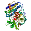
| ||||||||||||
|---|---|---|---|---|---|---|---|---|---|---|---|---|---|
| 1 |
| ||||||||||||
| Unit cell |
|
-Components
-Protein , 1 types, 1 molecules A
| #1: Protein | Mass: 55134.477 Da / Num. of mol.: 1 Source method: isolated from a genetically manipulated source Source: (gene. exp.) Geminocystis sp. (bacteria) / Strain: SKYG4 / Gene: ovoA, IGQ44_00985 / Production host: |
|---|
-Non-polymers , 5 types, 452 molecules 


| #2: Chemical | ChemComp-FE /  Mass: 55.845 Da / Num. of mol.: 1 / Source method: obtained synthetically / Formula: Fe Mass: 55.845 Da / Num. of mol.: 1 / Source method: obtained synthetically / Formula: Fe | ||||
|---|---|---|---|---|---|
| #3: Chemical | ChemComp-AVJ /  Mass: 198.242 Da / Num. of mol.: 1 / Source method: obtained synthetically / Formula: C9H16N3O2 / Feature type: SUBJECT OF INVESTIGATION Mass: 198.242 Da / Num. of mol.: 1 / Source method: obtained synthetically / Formula: C9H16N3O2 / Feature type: SUBJECT OF INVESTIGATION | ||||
| #4: Chemical | ChemComp-EDO /  Mass: 62.068 Da / Num. of mol.: 5 / Source method: obtained synthetically / Formula: C2H6O2 Mass: 62.068 Da / Num. of mol.: 5 / Source method: obtained synthetically / Formula: C2H6O2#5: Chemical | ChemComp-NA / |  Mass: 22.990 Da / Num. of mol.: 1 / Source method: obtained synthetically / Formula: Na Mass: 22.990 Da / Num. of mol.: 1 / Source method: obtained synthetically / Formula: Na#6: Water | ChemComp-HOH / | Mass: 18.015 Da / Num. of mol.: 444 / Source method: isolated from a natural source / Formula: H2O |
-Details
| Has ligand of interest | Y |
|---|---|
| Has protein modification | N |
-Experimental details
-Experiment
| Experiment | Method: X-RAY DIFFRACTION / Number of used crystals: 1 |
|---|
- Sample preparation
| Crystal | Density Matthews: 2.2 Å3/Da / Density % sol: 44 % / Description: Thin plates |
|---|---|
| Crystal grow | Temperature: 293 K / Method: vapor diffusion, sitting drop / pH: 6.7 Details: 0.1 M Bis-Tris, pH 6.7, 23% PEG3350, 0.2 M magnesium chloride |
-Data collection
| Diffraction | Mean temperature: 100 K / Serial crystal experiment: N |
|---|---|
| Diffraction source | Source: SYNCHROTRON / Site: CLSI  / Beamline: 08ID-1 / Wavelength: 0.9537 Å / Beamline: 08ID-1 / Wavelength: 0.9537 Å |
| Detector | Type: DECTRIS EIGER X 9M / Detector: PIXEL / Date: Aug 15, 2023 |
| Radiation | Protocol: SINGLE WAVELENGTH / Monochromatic (M) / Laue (L): M / Scattering type: x-ray |
| Radiation wavelength | Wavelength: 0.9537 Å / Relative weight: 1 |
| Reflection | Resolution: 1.6→47.82 Å / Num. obs: 61246 / % possible obs: 99.81 % / Redundancy: 2 % / Biso Wilson estimate: 19.52 Å2 / CC1/2: 0.996 / CC star: 0.999 / Rmerge(I) obs: 0.05404 / Rpim(I) all: 0.05404 / Rrim(I) all: 0.07642 / Net I/σ(I): 7.45 |
| Reflection shell | Resolution: 1.6→1.657 Å / Redundancy: 2 % / Rmerge(I) obs: 0.677 / Mean I/σ(I) obs: 1.02 / Num. unique obs: 5990 / CC1/2: 0.509 / CC star: 0.821 / Rpim(I) all: 0.677 / Rrim(I) all: 0.9574 / % possible all: 99.4 |
- Processing
| Software |
| |||||||||||||||||||||||||||||||||||||||||||||||||||||||||||||||||||||||||||||||||||||||||||||||||||||||||
|---|---|---|---|---|---|---|---|---|---|---|---|---|---|---|---|---|---|---|---|---|---|---|---|---|---|---|---|---|---|---|---|---|---|---|---|---|---|---|---|---|---|---|---|---|---|---|---|---|---|---|---|---|---|---|---|---|---|---|---|---|---|---|---|---|---|---|---|---|---|---|---|---|---|---|---|---|---|---|---|---|---|---|---|---|---|---|---|---|---|---|---|---|---|---|---|---|---|---|---|---|---|---|---|---|---|---|
| Refinement | Method to determine structure: MOLECULAR REPLACEMENT / Resolution: 1.6→47.82 Å / SU ML: 0.2339 / Cross valid method: FREE R-VALUE / σ(F): 1.34 / Phase error: 20.7325 Stereochemistry target values: GeoStd + Monomer Library + CDL v1.2
| |||||||||||||||||||||||||||||||||||||||||||||||||||||||||||||||||||||||||||||||||||||||||||||||||||||||||
| Solvent computation | Shrinkage radii: 0.9 Å / VDW probe radii: 1.1 Å / Solvent model: FLAT BULK SOLVENT MODEL | |||||||||||||||||||||||||||||||||||||||||||||||||||||||||||||||||||||||||||||||||||||||||||||||||||||||||
| Displacement parameters | Biso mean: 21.17 Å2 | |||||||||||||||||||||||||||||||||||||||||||||||||||||||||||||||||||||||||||||||||||||||||||||||||||||||||
| Refinement step | Cycle: LAST / Resolution: 1.6→47.82 Å
| |||||||||||||||||||||||||||||||||||||||||||||||||||||||||||||||||||||||||||||||||||||||||||||||||||||||||
| Refine LS restraints |
| |||||||||||||||||||||||||||||||||||||||||||||||||||||||||||||||||||||||||||||||||||||||||||||||||||||||||
| LS refinement shell |
|
