 Movie
Movie Controller


+ Open data

- Basic information
| Entry | Database: PDB / ID: 8vg6 | ||||||
|---|---|---|---|---|---|---|---|
| Title | Structure of Tulane virus | ||||||
 Components Components | Capsid protein | ||||||
 Keywords Keywords | VIRUS / Tualne virus / Calicivirus / norovirus | ||||||
| Function / homology | Calicivirus coat protein C-terminal / Calicivirus coat protein C-terminal / Calicivirus coat protein / Calicivirus coat protein / virion component / Viral coat protein subunit / host cell cytoplasm / Capsid protein Function and homology information Function and homology information | ||||||
| Biological species |  Tulane virus Tulane virus | ||||||
| Method | ELECTRON MICROSCOPY / single particle reconstruction / cryo EM / Resolution: 2.6 Å | ||||||
 Authors Authors | Yu, G. / Li, K. / Jiang, W. | ||||||
| Funding support |  United States, 1items United States, 1items
| ||||||
 Citation Citation | Journal: Structure / Year: 2016 Title: Antibody-Based Affinity Cryoelectron Microscopy at 2.6-Å Resolution. Authors: Guimei Yu / Kunpeng Li / Pengwei Huang / Xi Jiang / Wen Jiang / Abstract: The affinity cryoelectron microscopy (cryo-EM) approach has been explored in recent years to simplify and/or improve the sample preparation for cryo-EM, which can bring previously challenging ...The affinity cryoelectron microscopy (cryo-EM) approach has been explored in recent years to simplify and/or improve the sample preparation for cryo-EM, which can bring previously challenging specimens such as those of low abundance and/or unpurified ones within reach of the cryo-EM technique. Despite the demonstrated successes for solving structures to low to intermediate resolutions, the lack of near-atomic structures using this approach has led to a common perception of affinity cryo-EM as a niche technique incapable of reaching high resolutions. Here, we report a ∼2.6-Å structure solved using the antibody-based affinity grid approach with low-concentration Tulane virus purified from a low-yield cell-culture system that has been challenging to standard cryo-EM grid preparation. Quantitative analyses of the structure indicate data and reconstruction quality comparable with the conventional grid preparation method using samples at high concentration. | ||||||
| History |
|
- Structure visualization
| Structure viewer | Molecule: MolmilJmol/JSmol |
|---|
- Downloads & links
-Download
| PDBx/mmCIF format | 8vg6.cif.gz | 263.6 KB | Display | PDBx/mmCIF format |
|---|---|---|---|---|
| PDB format | pdb8vg6.ent.gz | 213.7 KB | Display | PDB format |
| PDBx/mmJSON format | 8vg6.json.gz | Tree view | PDBx/mmJSON format | |
| Others |  Other downloads Other downloads |
-Validation report
| Arichive directory | https://data.pdbj.org/pub/pdb/validation_reports/vg/8vg6ftp://data.pdbj.org/pub/pdb/validation_reports/vg/8vg6 | HTTPS FTP |
|---|
-Related structure data
| Related structure data | 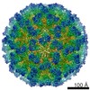 8252MC M: map data used to model this data C: citing same article ( |
|---|---|
| Similar structure data |
-Links
 PDBj
PDBj
- Assembly
| Deposited unit | 
|
|---|---|
| 1 | x 60
|
| 2 |
|
| 3 | x 5
|
| 4 | x 6
|
| 5 | 
|
| Symmetry | Point symmetry: (Schoenflies symbol: I (icosahedral)) |
-Components
| #1: Protein | Mass: 57890.055 Da / Num. of mol.: 3 Source method: isolated from a genetically manipulated source Source: (gene. exp.) Tulane virus / Plasmid: pBR322 / Cell line (production host): monkey kidney cells / Production host:  Platyrrhini (New World monkeys) / References: UniProt: B2Y6D0 Platyrrhini (New World monkeys) / References: UniProt: B2Y6D0Has protein modification | N | |
|---|
-Experimental details
-Experiment
| Experiment | Method: ELECTRON MICROSCOPY |
|---|---|
| EM experiment | Aggregation state: PARTICLE / 3D reconstruction method: single particle reconstruction |
- Sample preparation
| Component | Name: Tulane virus / Type: VIRUS / Details: Purified from cell cultures / Entity ID: all / Source: RECOMBINANT |
|---|---|
| Molecular weight | Value: 10 MDa / Experimental value: NO |
| Source (natural) | Organism: Tulane virus |
| Source (recombinant) | Organism: Platyrrhini (New World monkeys) / Cell: Monkey kidney cells / Plasmid: pBR322 |
| Details of virus | Empty: NO / Enveloped: NO / Isolate: STRAIN / Type: VIRION |
| Natural host | Organism: Macaca |
| Virus shell | Diameter: 400 nm |
| Buffer solution | pH: 7.4 |
| Specimen | Embedding applied: NO / Shadowing applied: NO / Staining applied: NO / Vitrification applied: YES |
| Vitrification | Cryogen name: ETHANE |
- Electron microscopy imaging
| Experimental equipment |  Model: Titan Krios / Image courtesy: FEI Company |
|---|---|
| Microscopy | Model: FEI TITAN KRIOS |
| Electron gun | Electron source:  FIELD EMISSION GUN / Accelerating voltage: 300 kV / Illumination mode: FLOOD BEAM FIELD EMISSION GUN / Accelerating voltage: 300 kV / Illumination mode: FLOOD BEAM |
| Electron lens | Mode: BRIGHT FIELD / Nominal defocus max: 3500 nm / Nominal defocus min: 500 nm |
| Image recording | Electron dose: 40 e/Å2 / Film or detector model: GATAN K2 SUMMIT (4k x 4k) |
- Processing
| EM software | Name: PHENIX / Category: model refinement | ||||||||||||||||||||||||
|---|---|---|---|---|---|---|---|---|---|---|---|---|---|---|---|---|---|---|---|---|---|---|---|---|---|
| CTF correction | Type: PHASE FLIPPING ONLY | ||||||||||||||||||||||||
| 3D reconstruction | Resolution: 2.6 Å / Resolution method: FSC 0.143 CUT-OFF / Num. of particles: 20000 / Symmetry type: POINT | ||||||||||||||||||||||||
| Refine LS restraints |
|
