 Movie
Movie Controller

[English] 日本語
 Yorodumi
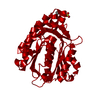
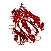
- PDB-8vdb: Crystal structure of Bacillus subtilis FabHB, beta-ketoacyl carri... -
+ Open data

- Basic information
| Entry | Database: PDB / ID: 8vdb | |||||||||
|---|---|---|---|---|---|---|---|---|---|---|
| Title | Crystal structure of Bacillus subtilis FabHB, beta-ketoacyl carrier protein synthase III | |||||||||
 Components Components | Beta-ketoacyl-[acyl-carrier-protein] synthase III 2 | |||||||||
 Keywords Keywords | TRANSFERASE / fatty acid biosynthesis / FASII / condensing enzyme | |||||||||
| Function / homology |  Function and homology information Function and homology informationbranched-chain beta-ketoacyl-[acyl-carrier-protein] synthase / beta-ketoacyl-[acyl-carrier-protein] synthase III / beta-ketoacyl-acyl-carrier-protein synthase III activity / secondary metabolite biosynthetic process / 3-oxoacyl-[acyl-carrier-protein] synthase activity / fatty acid biosynthetic process / cytoplasm Similarity search - Function | |||||||||
| Biological species |  | |||||||||
| Method |  X-RAY DIFFRACTION / SYNCHROTRON / MOLECULAR REPLACEMENT / Resolution: 2.4 Å X-RAY DIFFRACTION / SYNCHROTRON / MOLECULAR REPLACEMENT / Resolution: 2.4 Å | |||||||||
 Authors Authors | Radka, C.D. / Rock, C.O. | |||||||||
| Funding support |  United States, 2items United States, 2items
| |||||||||
 Citation Citation | Journal: J.Struct.Biol. / Year: 2024 Title: Crystal structures of the fatty acid biosynthesis initiation enzymes in Bacillus subtilis. Authors: Radka, C.D. / Rock, C.O. | |||||||||
| History |
|
- Structure visualization
| Structure viewer | Molecule: MolmilJmol/JSmol |
|---|
- Downloads & links
-Download
| PDBx/mmCIF format | 8vdb.cif.gz | 261.6 KB | Display | PDBx/mmCIF format |
|---|---|---|---|---|
| PDB format | pdb8vdb.ent.gz | 210.4 KB | Display | PDB format |
| PDBx/mmJSON format | 8vdb.json.gz | Tree view | PDBx/mmJSON format | |
| Others |  Other downloads Other downloads |
-Validation report
| Arichive directory | https://data.pdbj.org/pub/pdb/validation_reports/vd/8vdbftp://data.pdbj.org/pub/pdb/validation_reports/vd/8vdb | HTTPS FTP |
|---|
-Related structure data
-Links
 PDBj
PDBj

- Assembly
| Deposited unit | 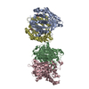
| ||||||||
|---|---|---|---|---|---|---|---|---|---|
| 1 | 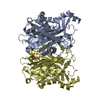
| ||||||||
| 2 | 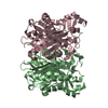
| ||||||||
| Unit cell |
|
-Components
| #1: Protein | Mass: 37635.648 Da / Num. of mol.: 4 Source method: isolated from a genetically manipulated source Source: (gene. exp.) References: UniProt: O07600, beta-ketoacyl-[acyl-carrier-protein] synthase III, branched-chain beta-ketoacyl-[acyl-carrier-protein] synthase #2: Chemical |   Mass: 92.094 Da / Num. of mol.: 3 / Source method: obtained synthetically / Formula: C3H8O3 Mass: 92.094 Da / Num. of mol.: 3 / Source method: obtained synthetically / Formula: C3H8O3#3: Water | ChemComp-HOH / |  Mass: 18.015 Da / Num. of mol.: 382 / Source method: isolated from a natural source / Formula: H2O Mass: 18.015 Da / Num. of mol.: 382 / Source method: isolated from a natural source / Formula: H2OHas ligand of interest | N | |
|---|
-Experimental details
-Experiment
| Experiment | Method: X-RAY DIFFRACTION / Number of used crystals: 1 |
|---|
- Sample preparation
| Crystal | Density Matthews: 4.15 Å3/Da / Density % sol: 70.34 % |
|---|---|
| Crystal grow | Temperature: 293.15 K / Method: vapor diffusion, hanging drop / Details: 20% MPD, 0.1 M HEPES, pH 6.5 |
-Data collection
| Diffraction | Mean temperature: 100 K / Serial crystal experiment: N |
|---|---|
| Diffraction source | Source: SYNCHROTRON / Site: APS / Beamline: 22-ID / Wavelength: 1 Å |
| Detector | Type: DECTRIS EIGER X 16M / Detector: PIXEL / Date: Jul 8, 2021 |
| Radiation | Protocol: SINGLE WAVELENGTH / Monochromatic (M) / Laue (L): M / Scattering type: x-ray |
| Radiation wavelength | Wavelength: 1 Å / Relative weight: 1 |
| Reflection | Resolution: 2.4→77.14 Å / Num. obs: 88179 / % possible obs: 93.2 % / Redundancy: 3.8 % / CC1/2: 0.983 / Rmerge(I) obs: 0.156 / Rrim(I) all: 0.182 / Net I/σ(I): 4.5 |
| Reflection shell | Resolution: 2.4→2.44 Å / Redundancy: 3.8 % / Rmerge(I) obs: 0.874 / Mean I/σ(I) obs: 1.8 / Num. unique obs: 4618 / CC1/2: 0.743 / % possible all: 94.1 |
- Processing
| Software |
| |||||||||||||||||||||||||||||||||||||||||||||||||||||||||||||||||||||||||||||||||||||||||||||||||||||||||
|---|---|---|---|---|---|---|---|---|---|---|---|---|---|---|---|---|---|---|---|---|---|---|---|---|---|---|---|---|---|---|---|---|---|---|---|---|---|---|---|---|---|---|---|---|---|---|---|---|---|---|---|---|---|---|---|---|---|---|---|---|---|---|---|---|---|---|---|---|---|---|---|---|---|---|---|---|---|---|---|---|---|---|---|---|---|---|---|---|---|---|---|---|---|---|---|---|---|---|---|---|---|---|---|---|---|---|
| Refinement | Method to determine structure: MOLECULAR REPLACEMENT / Resolution: 2.4→45.2 Å / SU ML: 0.26 / Cross valid method: THROUGHOUT / σ(F): 1.96 / Phase error: 27.43 / Stereochemistry target values: ML
| |||||||||||||||||||||||||||||||||||||||||||||||||||||||||||||||||||||||||||||||||||||||||||||||||||||||||
| Solvent computation | Shrinkage radii: 0.9 Å / VDW probe radii: 1.11 Å / Solvent model: FLAT BULK SOLVENT MODEL | |||||||||||||||||||||||||||||||||||||||||||||||||||||||||||||||||||||||||||||||||||||||||||||||||||||||||
| Refinement step | Cycle: LAST / Resolution: 2.4→45.2 Å
| |||||||||||||||||||||||||||||||||||||||||||||||||||||||||||||||||||||||||||||||||||||||||||||||||||||||||
| Refine LS restraints |
| |||||||||||||||||||||||||||||||||||||||||||||||||||||||||||||||||||||||||||||||||||||||||||||||||||||||||
| LS refinement shell |
|
