 Movie
Movie Controller

[English] 日本語
 Yorodumi
- PDB-8vc7: Crystal Structure of Human BTN2A1 ectodomain in complex with Anta... -
+ Open data

- Basic information
| Entry | Database: PDB / ID: 8vc7 | ||||||
|---|---|---|---|---|---|---|---|
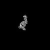
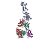
| Title | Crystal Structure of Human BTN2A1 ectodomain in complex with Antagonist 2A1.9 Fab | ||||||
 Components Components |
| ||||||
 Keywords Keywords | SIGNALING PROTEIN / Inhibitor / Complex / Antibody / Immune Recognition | ||||||
| Function / homology |  Function and homology information Function and homology informationButyrophilin (BTN) family interactions / regulation of cytokine production / lipid metabolic process / T cell receptor signaling pathway / external side of plasma membrane / signaling receptor binding / plasma membrane Similarity search - Function | ||||||
| Biological species |  Homo sapiens (human) Homo sapiens (human) | ||||||
| Method |  X-RAY DIFFRACTION / SYNCHROTRON / MOLECULAR REPLACEMENT / Resolution: 2.76 Å X-RAY DIFFRACTION / SYNCHROTRON / MOLECULAR REPLACEMENT / Resolution: 2.76 Å | ||||||
 Authors Authors | Ramesh, A. / Roy, S. / Adams, E. | ||||||
| Funding support |  United States, 1items United States, 1items
| ||||||
 Citation Citation | Journal: Sci Rep / Year: 2025 Title: Mapping the extracellular molecular architecture of the pAg-signaling complex with α-Butyrophilin antibodies. Authors: Amrita Ramesh / Sobhan Roy / Tomasz Slezak / James Fuller / Hortencia Graves / Murad R Mamedov / Alexander Marson / Anthony A Kossiakoff / Erin J Adams / Abstract: Target cells trigger Vγ9Vδ2 T cell activation by signaling the intracellular accumulation of phospho-antigen metabolites (pAgs) through Butyrophilin (BTN)-3A1 and BTN2A1 to the Vγ9Vδ2 T cell ...Target cells trigger Vγ9Vδ2 T cell activation by signaling the intracellular accumulation of phospho-antigen metabolites (pAgs) through Butyrophilin (BTN)-3A1 and BTN2A1 to the Vγ9Vδ2 T cell receptor (TCR). An incomplete understanding of the molecular dynamics in this signaling complex hampers Vγ9Vδ2 T cell immunotherapeutic efficacy. A panel of engineered α-BTN3A1 and α-BTN2A1 antibody (mAb) reagents was used to probe the roles of BTN3A1 and BTN2A1 in pAg signaling. Modified α-BTN3A1 mAbs with increased inter-Fab distances establish that tight clustering of BTN3A1 is not necessary to stimulate Vγ9Vδ2 T cell activation, and that antagonism may occur through occlusion of a critical binding interaction between BTN3A1 and a yet unknown co-receptor. Finally, a panel of additional α-BTN2A1 antagonists utilize different biophysical mechanisms to compete with Vγ9Vδ2 TCRs for BTN2A1 binding. The complex structures of BTN2A1 ectodomain and Fabs from three antagonist mAbs provide molecular insights into BTN2A1 epitopes critical for pAg-signaling. | ||||||
| History |
|
- Structure visualization
| Structure viewer | Molecule: MolmilJmol/JSmol |
|---|
- Downloads & links
-Download
| PDBx/mmCIF format | 8vc7.cif.gz | 255.2 KB | Display | PDBx/mmCIF format |
|---|---|---|---|---|
| PDB format | pdb8vc7.ent.gz | 202.7 KB | Display | PDB format |
| PDBx/mmJSON format | 8vc7.json.gz | Tree view | PDBx/mmJSON format | |
| Others |  Other downloads Other downloads |
-Validation report
| Arichive directory | https://data.pdbj.org/pub/pdb/validation_reports/vc/8vc7ftp://data.pdbj.org/pub/pdb/validation_reports/vc/8vc7 | HTTPS FTP |
|---|
-Related structure data
-Links
 PDBj
PDBj


- Assembly
| Deposited unit | 
| ||||||||
|---|---|---|---|---|---|---|---|---|---|
| 1 | 
| ||||||||
| 2 | 
| ||||||||
| Unit cell |
|
-Components
| #1: Antibody | Mass: 23258.783 Da / Num. of mol.: 2 Source method: isolated from a genetically manipulated source Details: Light chain of Herceptin Fab scaffold with CDR loops modified through phage-display evolution. (CDR1: VSSAV) (CDR2: IYSASSLY) (CDR3: SSSSLIT). N-terminal (S) residue is a linker/restriction ...Details: Light chain of Herceptin Fab scaffold with CDR loops modified through phage-display evolution. (CDR1: VSSAV) (CDR2: IYSASSLY) (CDR3: SSSSLIT). N-terminal (S) residue is a linker/restriction enzyme artifact, disordered and not modeled. Source: (gene. exp.) Homo sapiens (human) / Plasmid: RH2.2 / Production host:  #2: Antibody | Mass: 24836.652 Da / Num. of mol.: 2 Source method: isolated from a genetically manipulated source Details: Heavy chain of Herceptin Fab scaffold with CDR loops modified through phage-display evolution. (CDR1: NLYSSSI) (CDR2: YIYPSSGYTS) (CDR3: YYYTRGYPDGMDY). N-terminal (EISE) is a ...Details: Heavy chain of Herceptin Fab scaffold with CDR loops modified through phage-display evolution. (CDR1: NLYSSSI) (CDR2: YIYPSSGYTS) (CDR3: YYYTRGYPDGMDY). N-terminal (EISE) is a linker/restriction enzyme artifact and first residue of Fab, and disordered and not modeled. Middle (SSKSTSG) and C-terminal (KSCDKTHT) residues were disordered and not modeled. Source: (gene. exp.) Homo sapiens (human) / Plasmid: RH2.2 / Production host: #3: Protein | Mass: 25725.307 Da / Num. of mol.: 2 / Mutation: C219S Source method: isolated from a genetically manipulated source Details: BTN2A1 ectodomain with C219S mutation, and N- and C-terminal linkers. N-terminal (ADL) and C-terminal (VSPSGSGLEVLFQ) residues are disordered and not modeled. 3 glycans are denoted as NAG. Source: (gene. exp.) Homo sapiens (human) / Gene: BTN2A1, BT2.1, BTF1 / Plasmid: pAc-GP67a / Production host:  Trichoplusia ni (cabbage looper) / References: UniProt: Q7KYR7 Trichoplusia ni (cabbage looper) / References: UniProt: Q7KYR7#4: Sugar | ChemComp-NAG /   Type: D-saccharide, beta linking / Mass: 221.208 Da / Num. of mol.: 5 Type: D-saccharide, beta linking / Mass: 221.208 Da / Num. of mol.: 5Source method: isolated from a genetically manipulated source Formula: C8H15NO6 Has ligand of interest | N | Has protein modification | Y | |
|---|
-Experimental details
-Experiment
| Experiment | Method: X-RAY DIFFRACTION / Number of used crystals: 1 |
|---|
- Sample preparation
| Crystal | Density Matthews: 3.22 Å3/Da / Density % sol: 61.75 % / Description: Overlapping large plates |
|---|---|
| Crystal grow | Temperature: 298 K / Method: vapor diffusion, sitting drop / pH: 8.3 Details: Optimized well-G9 from Morpheus MIDAS MD1-76-HT Screen: - 0.1M Buffer System 3, pH 8.3 (Buffer System 3 1M Stock: 50.3% 1M Bicine, 49.7% 1M Tris) - 0.1M Carboxylic Acids Mix (Carboxylic ...Details: Optimized well-G9 from Morpheus MIDAS MD1-76-HT Screen: - 0.1M Buffer System 3, pH 8.3 (Buffer System 3 1M Stock: 50.3% 1M Bicine, 49.7% 1M Tris) - 0.1M Carboxylic Acids Mix (Carboxylic Acids Mix 1M Stock: 0.2M Sodium formate; 0.2M Ammonium acetate; 0.2M Sodium citrate tribasic dihydrate; 0.2M Potassium sodium tartrate tetrahydrate; 0.2M Sodium oxamate) - 20% Precipitant mix 1 (Precipitant mix 1 60% Stock: 40% v/v PEG500 MME; 20% w/v PEG20000) |
-Data collection
| Diffraction | Mean temperature: 100 K / Serial crystal experiment: N |
|---|---|
| Diffraction source | Source: SYNCHROTRON / Site: SSRL / Beamline: BL14-1 / Wavelength: 1.195 Å |
| Detector | Type: DECTRIS PILATUS 6M / Detector: PIXEL / Date: Aug 31, 2023 Details: Mirror: Flat bent collimating Rh coated mirror, toroidal focussing mirror |
| Radiation | Monochromator: Si (111) double crystal / Protocol: SINGLE WAVELENGTH / Monochromatic (M) / Laue (L): M / Scattering type: x-ray |
| Radiation wavelength | Wavelength: 1.195 Å / Relative weight: 1 |
| Reflection | Resolution: 2.76→39.46 Å / Num. obs: 49431 / % possible obs: 99.8 % / Redundancy: 8.9 % / Rmerge(I) obs: 0.258 / Rpim(I) all: 0.131 / Rrim(I) all: 0.29 / Net I/σ(I): 7.2 |
| Reflection shell | Resolution: 2.76→2.85 Å / Redundancy: 9.3 % / Rmerge(I) obs: 1.424 / Num. unique obs: 4503 / Rpim(I) all: 0.711 / Rrim(I) all: 1.595 / % possible all: 100 |
- Processing
| Software |
| |||||||||||||||||||||||||||||||||||||||||||||||||||||||||||||||||||||||||||||||||||||||||||||||||||||||||
|---|---|---|---|---|---|---|---|---|---|---|---|---|---|---|---|---|---|---|---|---|---|---|---|---|---|---|---|---|---|---|---|---|---|---|---|---|---|---|---|---|---|---|---|---|---|---|---|---|---|---|---|---|---|---|---|---|---|---|---|---|---|---|---|---|---|---|---|---|---|---|---|---|---|---|---|---|---|---|---|---|---|---|---|---|---|---|---|---|---|---|---|---|---|---|---|---|---|---|---|---|---|---|---|---|---|---|
| Refinement | Method to determine structure: MOLECULAR REPLACEMENT / Resolution: 2.76→39.46 Å / SU ML: 0.42 / Cross valid method: FREE R-VALUE / σ(F): 1.33 / Phase error: 27.33 / Stereochemistry target values: ML
| |||||||||||||||||||||||||||||||||||||||||||||||||||||||||||||||||||||||||||||||||||||||||||||||||||||||||
| Solvent computation | Shrinkage radii: 0.9 Å / VDW probe radii: 1.1 Å / Solvent model: FLAT BULK SOLVENT MODEL | |||||||||||||||||||||||||||||||||||||||||||||||||||||||||||||||||||||||||||||||||||||||||||||||||||||||||
| Refinement step | Cycle: LAST / Resolution: 2.76→39.46 Å
| |||||||||||||||||||||||||||||||||||||||||||||||||||||||||||||||||||||||||||||||||||||||||||||||||||||||||
| Refine LS restraints |
| |||||||||||||||||||||||||||||||||||||||||||||||||||||||||||||||||||||||||||||||||||||||||||||||||||||||||
| LS refinement shell |
|
