 Movie
Movie Controller


+ Open data

- Basic information
| Entry | Database: PDB / ID: 8vb1 | ||||||
|---|---|---|---|---|---|---|---|
| Title | Crystal structure of HIV-1 protease with GS-9770 | ||||||
 Components Components | HIV-1 protease | ||||||
 Keywords Keywords | VIRAL PROTEIN / HIV / protease / antiviral / inhibitor | ||||||
| Function / homology |  Function and homology information Function and homology information | ||||||
| Biological species |  HIV-1 06TG.HT043 (virus) HIV-1 06TG.HT043 (virus) | ||||||
| Method |  X-RAY DIFFRACTION / SYNCHROTRON / MOLECULAR REPLACEMENT / Resolution: 1.3 Å X-RAY DIFFRACTION / SYNCHROTRON / MOLECULAR REPLACEMENT / Resolution: 1.3 Å | ||||||
 Authors Authors | Lansdon, E.B. | ||||||
| Funding support | 1items
| ||||||
 Citation Citation | Journal: Antimicrob.Agents Chemother. / Year: 2024 Title: Preclinical characterization of a non-peptidomimetic HIV protease inhibitor with improved metabolic stability. Authors: Mulato, A. / Lansdon, E. / Aoyama, R. / Voigt, J. / Lee, M. / Liclican, A. / Lee, G. / Singer, E. / Stafford, B. / Gong, R. / Murray, B. / Chan, J. / Lee, J. / Xu, Y. / Ahmadyar, S. / ...Authors: Mulato, A. / Lansdon, E. / Aoyama, R. / Voigt, J. / Lee, M. / Liclican, A. / Lee, G. / Singer, E. / Stafford, B. / Gong, R. / Murray, B. / Chan, J. / Lee, J. / Xu, Y. / Ahmadyar, S. / Gonzalez, A. / Cho, A. / Stepan, G.J. / Schmitz, U. / Schultz, B. / Marchand, B. / Brumshtein, B. / Wang, R. / Yu, H. / Cihlar, T. / Xu, L. / Yant, S.R. | ||||||
| History |
|
- Structure visualization
| Structure viewer | Molecule: MolmilJmol/JSmol |
|---|
- Downloads & links
-Download
| PDBx/mmCIF format | 8vb1.cif.gz | 72.2 KB | Display | PDBx/mmCIF format |
|---|---|---|---|---|
| PDB format | pdb8vb1.ent.gz | Display | PDB format | |
| PDBx/mmJSON format | 8vb1.json.gz | Tree view | PDBx/mmJSON format | |
| Others |  Other downloads Other downloads |
-Validation report
| Summary document | 8vb1_validation.pdf.gz | 834.9 KB | Display | wwPDB validaton report |
|---|---|---|---|---|
| Full document | 8vb1_full_validation.pdf.gz | 836.4 KB | Display | |
| Data in XML | 8vb1_validation.xml.gz | 13 KB | Display | |
| Data in CIF | 8vb1_validation.cif.gz | 18.8 KB | Display | |
| Arichive directory | https://data.pdbj.org/pub/pdb/validation_reports/vb/8vb1ftp://data.pdbj.org/pub/pdb/validation_reports/vb/8vb1 | HTTPS FTP |
-Related structure data
| Similar structure data |
|---|
-Links
 PDBj
PDBj
- Assembly
| Deposited unit | 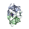
| ||||||||||||
|---|---|---|---|---|---|---|---|---|---|---|---|---|---|
| 1 |
| ||||||||||||
| Unit cell |
|
-Components
| #1: Protein | Mass: 10831.809 Da / Num. of mol.: 2 Source method: isolated from a genetically manipulated source Source: (gene. exp.) HIV-1 06TG.HT043 (virus) / Gene: pol / Production host:  #2: Chemical | ChemComp-A1AAD / ( | Mass: 811.151 Da / Num. of mol.: 1 / Source method: obtained synthetically / Formula: C35H34ClF7N10O3 / Feature type: SUBJECT OF INVESTIGATION #3: Water | ChemComp-HOH / |  Mass: 18.015 Da / Num. of mol.: 267 / Source method: isolated from a natural source / Formula: H2O Mass: 18.015 Da / Num. of mol.: 267 / Source method: isolated from a natural source / Formula: H2OHas ligand of interest | Y | |
|---|
-Experimental details
-Experiment
| Experiment | Method: X-RAY DIFFRACTION / Number of used crystals: 1 |
|---|
- Sample preparation
| Crystal | Density Matthews: 2.62 Å3/Da / Density % sol: 53.08 % |
|---|---|
| Crystal grow | Temperature: 293 K / Method: vapor diffusion, hanging drop / pH: 6.5 / Details: 2.25M ammonium acetate 100mM MES pH 6.5 |
-Data collection
| Diffraction | Mean temperature: 170 K / Serial crystal experiment: N |
|---|---|
| Diffraction source | Source: SYNCHROTRON / Site: ALS  / Beamline: 5.0.1 / Wavelength: 1 Å / Beamline: 5.0.1 / Wavelength: 1 Å |
| Detector | Type: DECTRIS PILATUS 6M / Detector: PIXEL / Date: May 4, 2017 |
| Radiation | Protocol: SINGLE WAVELENGTH / Monochromatic (M) / Laue (L): M / Scattering type: x-ray |
| Radiation wavelength | Wavelength: 1 Å / Relative weight: 1 |
| Reflection | Resolution: 1.3→47.5 Å / Num. obs: 54560 / % possible obs: 99.7 % / Redundancy: 7.2 % / Biso Wilson estimate: 12.65 Å2 / CC1/2: 0.999 / Net I/σ(I): 16.8 |
| Reflection shell | Resolution: 1.3→1.38 Å / Num. unique obs: 4483 / CC1/2: 0.722 |
- Processing
| Software |
| |||||||||||||||||||||||||||||||||||||||||||||||||||||||||||||||||||||||||||||||||||||||||||||||||||||||||
|---|---|---|---|---|---|---|---|---|---|---|---|---|---|---|---|---|---|---|---|---|---|---|---|---|---|---|---|---|---|---|---|---|---|---|---|---|---|---|---|---|---|---|---|---|---|---|---|---|---|---|---|---|---|---|---|---|---|---|---|---|---|---|---|---|---|---|---|---|---|---|---|---|---|---|---|---|---|---|---|---|---|---|---|---|---|---|---|---|---|---|---|---|---|---|---|---|---|---|---|---|---|---|---|---|---|---|
| Refinement | Method to determine structure: MOLECULAR REPLACEMENT / Resolution: 1.3→47.47 Å / SU ML: 0.1518 / Cross valid method: FREE R-VALUE / σ(F): 0.08 / Phase error: 22.1071 Stereochemistry target values: GeoStd + Monomer Library + CDL v1.2
| |||||||||||||||||||||||||||||||||||||||||||||||||||||||||||||||||||||||||||||||||||||||||||||||||||||||||
| Solvent computation | Shrinkage radii: 0.9 Å / VDW probe radii: 1.1 Å / Solvent model: FLAT BULK SOLVENT MODEL | |||||||||||||||||||||||||||||||||||||||||||||||||||||||||||||||||||||||||||||||||||||||||||||||||||||||||
| Displacement parameters | Biso mean: 17.22 Å2 | |||||||||||||||||||||||||||||||||||||||||||||||||||||||||||||||||||||||||||||||||||||||||||||||||||||||||
| Refinement step | Cycle: LAST / Resolution: 1.3→47.47 Å
| |||||||||||||||||||||||||||||||||||||||||||||||||||||||||||||||||||||||||||||||||||||||||||||||||||||||||
| Refine LS restraints |
| |||||||||||||||||||||||||||||||||||||||||||||||||||||||||||||||||||||||||||||||||||||||||||||||||||||||||
| LS refinement shell |
|
