 Movie
Movie Controller

[English] 日本語
 Yorodumi
- PDB-8v2y: Room temperature X-ray Crystal Structure of FMN-bound long-chain ... -
+ Open data

- Basic information
| Entry | Database: PDB / ID: 8v2y | ||||||
|---|---|---|---|---|---|---|---|
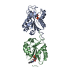
| Title | Room temperature X-ray Crystal Structure of FMN-bound long-chain flavodoxin from Rhodopseudomonas palustris | ||||||
 Components Components | Flavodoxin | ||||||
 Keywords Keywords | FLAVOPROTEIN / FMN-bound long chain Flavodoxin | ||||||
| Function / homology |  Function and homology information Function and homology information | ||||||
| Biological species |  Rhodopseudomonas palustris (phototrophic) Rhodopseudomonas palustris (phototrophic) | ||||||
| Method |  X-RAY DIFFRACTION / MOLECULAR REPLACEMENT / Resolution: 2.86 Å X-RAY DIFFRACTION / MOLECULAR REPLACEMENT / Resolution: 2.86 Å | ||||||
 Authors Authors | Ansari, A. / Khan, S.A. / Miller, A.F. | ||||||
| Funding support |  United States, 1items United States, 1items
| ||||||
 Citation Citation | Journal: J.Biol.Chem. / Year: 2024 Title: Structure, dynamics, and redox reactivity of an all-purpose flavodoxin. Authors: Khan, S. / Ansari, A. / Brachi, M. / Das, D. / El Housseini, W. / Minteer, S. / Miller, A.F. | ||||||
| History |
|
- Structure visualization
| Structure viewer | Molecule: MolmilJmol/JSmol |
|---|
- Downloads & links
-Download
| PDBx/mmCIF format | 8v2y.cif.gz | 82.7 KB | Display | PDBx/mmCIF format |
|---|---|---|---|---|
| PDB format | pdb8v2y.ent.gz | 54.5 KB | Display | PDB format |
| PDBx/mmJSON format | 8v2y.json.gz | Tree view | PDBx/mmJSON format | |
| Others |  Other downloads Other downloads |
-Validation report
| Summary document | 8v2y_validation.pdf.gz | 956.3 KB | Display | wwPDB validaton report |
|---|---|---|---|---|
| Full document | 8v2y_full_validation.pdf.gz | 959 KB | Display | |
| Data in XML | 8v2y_validation.xml.gz | 14.2 KB | Display | |
| Data in CIF | 8v2y_validation.cif.gz | 19 KB | Display | |
| Arichive directory | https://data.pdbj.org/pub/pdb/validation_reports/v2/8v2yftp://data.pdbj.org/pub/pdb/validation_reports/v2/8v2y | HTTPS FTP |
-Related structure data
-Links
 PDBj
PDBj

- Assembly
| Deposited unit | 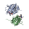
| |||||||||||||||||||||||||||||||||||||||||||||||||||||||||||||||||||||||||||||||||||||||||||||||||||||||||||||
|---|---|---|---|---|---|---|---|---|---|---|---|---|---|---|---|---|---|---|---|---|---|---|---|---|---|---|---|---|---|---|---|---|---|---|---|---|---|---|---|---|---|---|---|---|---|---|---|---|---|---|---|---|---|---|---|---|---|---|---|---|---|---|---|---|---|---|---|---|---|---|---|---|---|---|---|---|---|---|---|---|---|---|---|---|---|---|---|---|---|---|---|---|---|---|---|---|---|---|---|---|---|---|---|---|---|---|---|---|---|---|
| 1 | 
| |||||||||||||||||||||||||||||||||||||||||||||||||||||||||||||||||||||||||||||||||||||||||||||||||||||||||||||
| Unit cell |
| |||||||||||||||||||||||||||||||||||||||||||||||||||||||||||||||||||||||||||||||||||||||||||||||||||||||||||||
| Noncrystallographic symmetry (NCS) | NCS domain:
NCS domain segments: Ens-ID: ens_1
NCS oper: (Code: givenMatrix: (-0.995035782273, 0.0075197324557, -0.0992332888701), (0.0366232366679, 0.954833993936, -0.294874180896), (0.0925339425948, -0.297044605484, -0.950369387039)Vector: 0. ...NCS oper: (Code: given Matrix: (-0.995035782273, 0.0075197324557, -0.0992332888701), Vector: |
-Components
| #1: Protein | Mass: 17884.822 Da / Num. of mol.: 2 Source method: isolated from a genetically manipulated source Source: (gene. exp.) Rhodopseudomonas palustris (phototrophic)Gene: RPA2117 / Production host: #2: Chemical |   Mass: 456.344 Da / Num. of mol.: 2 / Source method: obtained synthetically / Formula: C17H21N4O9P / Feature type: SUBJECT OF INVESTIGATION Mass: 456.344 Da / Num. of mol.: 2 / Source method: obtained synthetically / Formula: C17H21N4O9P / Feature type: SUBJECT OF INVESTIGATION#3: Water | ChemComp-HOH / |  Mass: 18.015 Da / Num. of mol.: 86 / Source method: isolated from a natural source / Formula: H2O Mass: 18.015 Da / Num. of mol.: 86 / Source method: isolated from a natural source / Formula: H2OHas ligand of interest | Y | |
|---|
-Experimental details
-Experiment
| Experiment | Method: X-RAY DIFFRACTION / Number of used crystals: 1 |
|---|
- Sample preparation
| Crystal | Density Matthews: 2.26 Å3/Da / Density % sol: 45.46 % |
|---|---|
| Crystal grow | Temperature: 287 K / Method: vapor diffusion, hanging drop / pH: 7.5 Details: 0.3-0.6M magnesium formate, 15-20% polyethylene glycol (PEG) 3350 |
-Data collection
| Diffraction | Mean temperature: 293 K / Serial crystal experiment: N |
|---|---|
| Diffraction source | Source: ROTATING ANODE / Type: RIGAKU / Wavelength: 1.5406 Å |
| Detector | Type: DECTRIS EIGER R 4M / Detector: PIXEL / Date: Oct 18, 2021 |
| Radiation | Protocol: SINGLE WAVELENGTH / Monochromatic (M) / Laue (L): M / Scattering type: x-ray |
| Radiation wavelength | Wavelength: 1.5406 Å / Relative weight: 1 |
| Reflection | Resolution: 2.86→28.27 Å / Num. obs: 7368 / % possible obs: 97.83 % / Redundancy: 3.6 % / Biso Wilson estimate: 47.92 Å2 / CC1/2: 0.922 / Rmerge(I) obs: 0.1291 / Rrim(I) all: 0.1528 / Net I/σ(I): 9.11 |
| Reflection shell | Resolution: 2.86→3.08 Å / Num. unique obs: 1366 / CC1/2: 0.842 |
- Processing
| Software |
| ||||||||||||||||||||||||||||||||||||||||||
|---|---|---|---|---|---|---|---|---|---|---|---|---|---|---|---|---|---|---|---|---|---|---|---|---|---|---|---|---|---|---|---|---|---|---|---|---|---|---|---|---|---|---|---|
| Refinement | Method to determine structure: MOLECULAR REPLACEMENT / Resolution: 2.86→28.27 Å / SU ML: 0.3659 / Cross valid method: FREE R-VALUE / σ(F): 1.37 / Phase error: 28.1821 Stereochemistry target values: GeoStd + Monomer Library + CDL v1.2
| ||||||||||||||||||||||||||||||||||||||||||
| Solvent computation | Shrinkage radii: 0.9 Å / VDW probe radii: 1.1 Å / Solvent model: FLAT BULK SOLVENT MODEL | ||||||||||||||||||||||||||||||||||||||||||
| Displacement parameters | Biso mean: 48.74 Å2 | ||||||||||||||||||||||||||||||||||||||||||
| Refinement step | Cycle: LAST / Resolution: 2.86→28.27 Å
| ||||||||||||||||||||||||||||||||||||||||||
| Refine LS restraints |
| ||||||||||||||||||||||||||||||||||||||||||
| Refine LS restraints NCS | Type: Torsion NCS / Rms dev position: 0.735412693386 Å | ||||||||||||||||||||||||||||||||||||||||||
| LS refinement shell |
|
