Movie
Movie Controller


+ Open data

- Basic information
| Entry | Database: PDB / ID: 8v2i | ||||||
|---|---|---|---|---|---|---|---|
| Title | Structure of alpha1B and betaI/IVb microtubule bound to GMPCPP | ||||||
 Components Components |
| ||||||
 Keywords Keywords |  STRUCTURAL PROTEIN / human microtubule / tubulin isoforms / cytoskeleton STRUCTURAL PROTEIN / human microtubule / tubulin isoforms / cytoskeleton | ||||||
| Function / homology |  Function and homology information Function and homology informationodontoblast differentiation / Post-chaperonin tubulin folding pathway / Carboxyterminal post-translational modifications of tubulin / Microtubule-dependent trafficking of connexons from Golgi to the plasma membrane / Cilium Assembly / cytoskeleton-dependent intracellular transport / Intraflagellar transport / Sealing of the nuclear envelope (NE) by ESCRT-III / Gap junction assembly / Formation of tubulin folding intermediates by CCT/TriC ...odontoblast differentiation / Post-chaperonin tubulin folding pathway / Carboxyterminal post-translational modifications of tubulin / Microtubule-dependent trafficking of connexons from Golgi to the plasma membrane / Cilium Assembly / cytoskeleton-dependent intracellular transport / Intraflagellar transport / Sealing of the nuclear envelope (NE) by ESCRT-III / Gap junction assembly / Formation of tubulin folding intermediates by CCT/TriC / natural killer cell mediated cytotoxicity / COPI-independent Golgi-to-ER retrograde traffic / Kinesins / Assembly and cell surface presentation of NMDA receptors / GTPase activating protein binding / COPI-dependent Golgi-to-ER retrograde traffic / regulation of synapse organization / intercellular bridge / nuclear envelope lumen / Recycling pathway of L1 / RHOH GTPase cycle / spindle assembly / RHO GTPases activate IQGAPs / MHC class I protein binding / Hedgehog 'off' state / cytoplasmic microtubule / microtubule-based process / COPI-mediated anterograde transport / Activation of AMPK downstream of NMDARs / Mitotic Prometaphase / EML4 and NUDC in mitotic spindle formation / Loss of Nlp from mitotic centrosomes / Loss of proteins required for interphase microtubule organization from the centrosome / Recruitment of mitotic centrosome proteins and complexes / Resolution of Sister Chromatid Cohesion / Recruitment of NuMA to mitotic centrosomes / HSP90 chaperone cycle for steroid hormone receptors (SHR) in the presence of ligand / Anchoring of the basal body to the plasma membrane / MHC class II antigen presentation / cellular response to interleukin-4 / AURKA Activation by TPX2 / RHO GTPases Activate Formins / Translocation of SLC2A4 (GLUT4) to the plasma membrane / Hydrolases; Acting on acid anhydrides; Acting on GTP to facilitate cellular and subcellular movement / PKR-mediated signaling / structural constituent of cytoskeleton / cytoplasmic ribonucleoprotein granule / mitotic spindle / microtubule cytoskeleton organization / Aggrephagy / HCMV Early Events / Separation of Sister Chromatids / The role of GTSE1 in G2/M progression after G2 checkpoint / microtubule cytoskeleton / azurophil granule lumen / Regulation of PLK1 Activity at G2/M Transition / double-stranded RNA binding / mitotic cell cycle / cell body / microtubule / Potential therapeutics for SARS / cytoskeleton / membrane raft / cell division / protein domain specific binding / GTPase activity / ubiquitin protein ligase binding / Neutrophil degranulation / protein-containing complex binding / GTP binding / structural molecule activity / protein-containing complex / extracellular exosome / extracellular region / metal ion binding / nucleus / cytosol / cytoplasmSimilarity search - Function | ||||||
| Biological species |  Homo sapiens (human) Homo sapiens (human) | ||||||
| Method | ELECTRON MICROSCOPY / single particle reconstruction / cryo EM / Resolution: 2.9 Å | ||||||
 Authors Authors | Zehr, E.A. / Roll-Mecak, A. | ||||||
| Funding support |  United States, 1items United States, 1items
| ||||||
 Citation Citation | Journal: Biorxiv / Year: 2023 Title: Cryo-EM structures of human a1B/bI+bIVb microtubules shed light on isoform specific assembly Authors: Zehr, E.A. / Roll-Mecak, A. | ||||||
| History |
|
- Structure visualization
| Structure viewer | Molecule: MolmilJmol/JSmol |
|---|
- Downloads & links
-Download
| PDBx/mmCIF format | 8v2i.cif.gz | 346.5 KB | Display | PDBx/mmCIF format |
|---|---|---|---|---|
| PDB format | pdb8v2i.ent.gz | 276.1 KB | Display | PDB format |
| PDBx/mmJSON format | 8v2i.json.gz | Tree view | PDBx/mmJSON format | |
| Others |  Other downloads Other downloads |
-Validation report
| Arichive directory | https://data.pdbj.org/pub/pdb/validation_reports/v2/8v2iftp://data.pdbj.org/pub/pdb/validation_reports/v2/8v2i | HTTPS FTP |
|---|
-Related structure data
| Related structure data | 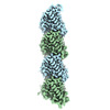 42915MC  42916 8v2j M: map data used to model this data C: citing same article ( |
|---|---|
| Similar structure data |
-Links
 PDBj
PDBj


- Assembly
| Deposited unit | 
|
|---|---|
| 1 |
|
-Components
| #1: Protein | Mass: 50204.445 Da / Num. of mol.: 2 / Source method: isolated from a natural source / Source: (natural) Homo sapiens (human) / Cell line: tsA201 / Organ: kidney / References: UniProt: P68363#2: Protein | Mass: 49717.629 Da / Num. of mol.: 2 / Source method: isolated from a natural source / Source: (natural) Homo sapiens (human) / Cell line: tsA201 / Organ: kidney / References: UniProt: P07437#3: Chemical | Guanosine triphosphate  Mass: 523.180 Da / Num. of mol.: 2 / Source method: obtained synthetically / Formula: C10H16N5O14P3 / Comment: GTP, energy-carrying molecule*YM Mass: 523.180 Da / Num. of mol.: 2 / Source method: obtained synthetically / Formula: C10H16N5O14P3 / Comment: GTP, energy-carrying molecule*YM#4: Chemical | ChemComp-MG /   Mass: 24.305 Da / Num. of mol.: 4 / Source method: obtained synthetically / Formula: Mg Mass: 24.305 Da / Num. of mol.: 4 / Source method: obtained synthetically / Formula: Mg#5: Chemical |   Mass: 521.208 Da / Num. of mol.: 2 / Source method: obtained synthetically / Formula: C11H18N5O13P3 / Feature type: SUBJECT OF INVESTIGATION / Comment: GMP-CPP, energy-carrying molecule analogue*YM Mass: 521.208 Da / Num. of mol.: 2 / Source method: obtained synthetically / Formula: C11H18N5O13P3 / Feature type: SUBJECT OF INVESTIGATION / Comment: GMP-CPP, energy-carrying molecule analogue*YMHas ligand of interest | Y | |
|---|
-Experimental details
-Experiment
| Experiment | Method: ELECTRON MICROSCOPY |
|---|---|
| EM experiment | Aggregation state: FILAMENT / 3D reconstruction method: single particle reconstruction |
- Sample preparation
| Component | Name: alpha1B and betaI/IVb microtubule / Type: COMPLEX / Details: Microtubules were double-cycled with GMPCPP / Entity ID: #1-#2 / Source: NATURAL | |||||||||||||||||||||||||
|---|---|---|---|---|---|---|---|---|---|---|---|---|---|---|---|---|---|---|---|---|---|---|---|---|---|---|
| Molecular weight | Value: 0.3 MDa / Experimental value: YES | |||||||||||||||||||||||||
| Source (natural) | Organism: Homo sapiens (human) / Cellular location: cytoplasm / Organ: kidney | |||||||||||||||||||||||||
| Buffer solution | pH: 7 | |||||||||||||||||||||||||
| Buffer component |
| |||||||||||||||||||||||||
| Specimen | Conc.: 0.1 mg/ml / Embedding applied: NO / Shadowing applied: NO / Staining applied: NO / Vitrification applied: YES Details: Microtubules were double-cycled with GMPCPP at 2mg/ml | |||||||||||||||||||||||||
| Specimen support | Grid material: GOLD / Grid mesh size: 300 divisions/in. / Grid type: C-flat-1.2/1.3 | |||||||||||||||||||||||||
| Vitrification | Instrument: LEICA EM GP / Cryogen name: ETHANE / Humidity: 100 % / Chamber temperature: 303 K Details: Double-cycled GMPCPP microtubules were diluted to 2.5uM in Brb80 buffer. 5 ul of microtubules were applied to glow-discharged cryo-EM grid and allowed to absorb for 30 sec. |
- Electron microscopy imaging
| Experimental equipment |  Model: Titan Krios / Image courtesy: FEI Company |
|---|---|
| Microscopy | Model: FEI TITAN KRIOS |
| Electron gun | Electron source: FIELD EMISSION GUN / Accelerating voltage: 300 kV / Illumination mode: FLOOD BEAM |
| Electron lens | Mode: BRIGHT FIELDBright-field microscopy / Nominal magnification: 130000 X / Nominal defocus max: 2500 nm / Nominal defocus min: 500 nm / Cs: 2.7 mm / Alignment procedure: COMA FREE |
| Image recording | Average exposure time: 7.8 sec. / Electron dose: 56 e/Å2 / Detector mode: COUNTING / Film or detector model: GATAN K2 SUMMIT (4k x 4k) / Num. of real images: 6930 |
| EM imaging optics | Energyfilter name: GIF Bioquantum / Energyfilter slit width: 20 eV |
| Image scans | Movie frames/image: 40 / Used frames/image: 1-40 |
- Processing
| EM software |
| ||||||||||||||||||||||||||||||||||||
|---|---|---|---|---|---|---|---|---|---|---|---|---|---|---|---|---|---|---|---|---|---|---|---|---|---|---|---|---|---|---|---|---|---|---|---|---|---|
| CTF correction | Type: PHASE FLIPPING AND AMPLITUDE CORRECTION | ||||||||||||||||||||||||||||||||||||
| Particle selection | Num. of particles selected: 124408 / Details: Manual picking | ||||||||||||||||||||||||||||||||||||
| Symmetry | Point symmetry: C1 (asymmetric) | ||||||||||||||||||||||||||||||||||||
| 3D reconstruction | Resolution: 2.9 Å / Resolution method: FSC 0.143 CUT-OFF / Num. of particles: 1293027 / Symmetry type: POINT | ||||||||||||||||||||||||||||||||||||
| Atomic model building | Protocol: FLEXIBLE FIT / Space: REAL / Target criteria: Cross-correlation | ||||||||||||||||||||||||||||||||||||
| Atomic model building | PDB-ID: 5N5N Accession code: 5N5N / Chain residue range: 1-441 / Pdb chain residue range: 1-441 / Source name: PDB / Type: experimental model |