 Movie
Movie Controller


+ Open data

- Basic information
| Entry | Database: PDB / ID: 8v14 | |||||||||
|---|---|---|---|---|---|---|---|---|---|---|
| Title | Structure of NRAP-1 and its role in NMDAR signaling | |||||||||
 Components Components | NMDA receptor auxiliary protein | |||||||||
 Keywords Keywords | SIGNALING PROTEIN / NMDAR / NRAP-1 / Synapse / Postsynaptic / Neurotransmitter | |||||||||
| Function / homology | Low-density lipoprotein receptor domain class A / LDL-receptor class A (LDLRA) domain profile. / Low-density lipoprotein receptor domain class A / Low-density lipoprotein (LDL) receptor class A repeat / LDL receptor-like superfamily / NMDA receptor auxiliary protein Function and homology information Function and homology information | |||||||||
| Biological species |  | |||||||||
| Method |  X-RAY DIFFRACTION / SYNCHROTRON / SAD / Resolution: 1.9 Å X-RAY DIFFRACTION / SYNCHROTRON / SAD / Resolution: 1.9 Å | |||||||||
 Authors Authors | Whitby, F.G. / Goodell, D.J. / Maricq, A.V. / Hill, C.P. | |||||||||
| Funding support |  United States, 2items United States, 2items
| |||||||||
 Citation Citation | Journal: To Be Published Title: Functional and Structural Studies of NRAP-1 and NMDAR signaling Authors: Goodell, D.M. / Maricq, A.V. | |||||||||
| History |
|
- Structure visualization
| Structure viewer | Molecule: MolmilJmol/JSmol |
|---|
- Downloads & links
-Download
| PDBx/mmCIF format | 8v14.cif.gz | 146.9 KB | Display | PDBx/mmCIF format |
|---|---|---|---|---|
| PDB format | pdb8v14.ent.gz | 115.4 KB | Display | PDB format |
| PDBx/mmJSON format | 8v14.json.gz | Tree view | PDBx/mmJSON format | |
| Others |  Other downloads Other downloads |
-Validation report
| Summary document | 8v14_validation.pdf.gz | 1.3 MB | Display | wwPDB validaton report |
|---|---|---|---|---|
| Full document | 8v14_full_validation.pdf.gz | 1.3 MB | Display | |
| Data in XML | 8v14_validation.xml.gz | 16.4 KB | Display | |
| Data in CIF | 8v14_validation.cif.gz | 23.3 KB | Display | |
| Arichive directory | https://data.pdbj.org/pub/pdb/validation_reports/v1/8v14ftp://data.pdbj.org/pub/pdb/validation_reports/v1/8v14 | HTTPS FTP |
-Related structure data
| Similar structure data |
|---|
-Links
 PDBj
PDBj
- Assembly
| Deposited unit | 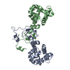
| ||||||||
|---|---|---|---|---|---|---|---|---|---|
| 1 |
| ||||||||
| Unit cell |
|
-Components
| #1: Protein | Mass: 18155.447 Da / Num. of mol.: 2 Source method: isolated from a genetically manipulated source Source: (gene. exp.)  Homo sapiens (human) / References: UniProt: G5EEF9 Homo sapiens (human) / References: UniProt: G5EEF9#2: Chemical |   Mass: 40.078 Da / Num. of mol.: 2 / Source method: obtained synthetically / Formula: Ca / Feature type: SUBJECT OF INVESTIGATION Mass: 40.078 Da / Num. of mol.: 2 / Source method: obtained synthetically / Formula: Ca / Feature type: SUBJECT OF INVESTIGATION#3: Water | ChemComp-HOH / |  Mass: 18.015 Da / Num. of mol.: 193 / Source method: isolated from a natural source / Formula: H2O Mass: 18.015 Da / Num. of mol.: 193 / Source method: isolated from a natural source / Formula: H2OHas ligand of interest | Y | |
|---|
-Experimental details
-Experiment
| Experiment | Method: X-RAY DIFFRACTION / Number of used crystals: 1 |
|---|
- Sample preparation
| Crystal | Density Matthews: 3.29 Å3/Da / Density % sol: 62.66 % |
|---|---|
| Crystal grow | Temperature: 277 K / Method: vapor diffusion, sitting drop / pH: 7.35 Details: 17 mg/mL purified NRAP-1 in 150 mM sodium chloride, 15 mM HEPES, 1 mM calcium chloride, pH 7.35, 1:1 with crystallization solution (26% PEG8000, 100 mM CHES, pH 9.8) |
-Data collection
| Diffraction | Mean temperature: 100 K / Serial crystal experiment: N | ||||||||||||||||||||||||||||||
|---|---|---|---|---|---|---|---|---|---|---|---|---|---|---|---|---|---|---|---|---|---|---|---|---|---|---|---|---|---|---|---|
| Diffraction source | Source: SYNCHROTRON / Site: SSRL / Beamline: BL9-2 / Wavelength: 1 Å | ||||||||||||||||||||||||||||||
| Detector | Type: DECTRIS PILATUS 6M / Detector: PIXEL / Date: Mar 5, 2020 | ||||||||||||||||||||||||||||||
| Radiation | Protocol: SINGLE WAVELENGTH / Monochromatic (M) / Laue (L): M / Scattering type: x-ray | ||||||||||||||||||||||||||||||
| Radiation wavelength | Wavelength: 1 Å / Relative weight: 1 | ||||||||||||||||||||||||||||||
| Reflection | Resolution: 1.9→38.12 Å / Num. obs: 38804 / % possible obs: 100 % / Redundancy: 54.8 % / CC1/2: 1 / Rmerge(I) obs: 0.111 / Rpim(I) all: 0.015 / Rrim(I) all: 0.112 / Net I/σ(I): 27.4 | ||||||||||||||||||||||||||||||
| Reflection shell | Diffraction-ID: 1
|
- Processing
| Software |
| |||||||||||||||||||||||||||||||||||||||||||||||||||||||||||||||||||||||||||
|---|---|---|---|---|---|---|---|---|---|---|---|---|---|---|---|---|---|---|---|---|---|---|---|---|---|---|---|---|---|---|---|---|---|---|---|---|---|---|---|---|---|---|---|---|---|---|---|---|---|---|---|---|---|---|---|---|---|---|---|---|---|---|---|---|---|---|---|---|---|---|---|---|---|---|---|---|
| Refinement | Method to determine structure: SAD / Resolution: 1.9→38.12 Å / Cor.coef. Fo:Fc: 0.966 / Cor.coef. Fo:Fc free: 0.955 / SU B: 7.106 / SU ML: 0.101 / SU R Cruickshank DPI: 0.1226 / Cross valid method: THROUGHOUT / σ(F): 0 / ESU R: 0.123 / ESU R Free: 0.121 / Stereochemistry target values: MAXIMUM LIKELIHOOD Details: HYDROGENS HAVE BEEN ADDED IN THE RIDING POSITIONS U VALUES : WITH TLS ADDED
| |||||||||||||||||||||||||||||||||||||||||||||||||||||||||||||||||||||||||||
| Solvent computation | Ion probe radii: 0.8 Å / Shrinkage radii: 0.8 Å / VDW probe radii: 1.2 Å / Solvent model: MASK | |||||||||||||||||||||||||||||||||||||||||||||||||||||||||||||||||||||||||||
| Displacement parameters | Biso max: 194.44 Å2 / Biso mean: 52.871 Å2 / Biso min: 28.65 Å2
| |||||||||||||||||||||||||||||||||||||||||||||||||||||||||||||||||||||||||||
| Refinement step | Cycle: final / Resolution: 1.9→38.12 Å
| |||||||||||||||||||||||||||||||||||||||||||||||||||||||||||||||||||||||||||
| Refine LS restraints |
| |||||||||||||||||||||||||||||||||||||||||||||||||||||||||||||||||||||||||||
| LS refinement shell | Resolution: 1.901→1.95 Å / Rfactor Rfree error: 0 / Total num. of bins used: 20
| |||||||||||||||||||||||||||||||||||||||||||||||||||||||||||||||||||||||||||
| Refinement TLS params. | Method: refined / Refine-ID: X-RAY DIFFRACTION
| |||||||||||||||||||||||||||||||||||||||||||||||||||||||||||||||||||||||||||
| Refinement TLS group |
|
