 Movie
Movie Controller

[English] 日本語
 Yorodumi
- PDB-8uz8: Crystal Structure of CiaD from Campylobacter jejuni (C-terminal f... -
+ Open data

- Basic information
| Entry | Database: PDB / ID: 8uz8 | |||||||||
|---|---|---|---|---|---|---|---|---|---|---|
| Title | Crystal Structure of CiaD from Campylobacter jejuni (C-terminal fragment, Orthorhombic P form) | |||||||||
 Components Components | 2-oxoglutarate:acceptor oxidoreductase | |||||||||
 Keywords Keywords | OXIDOREDUCTASE / SSGCID / STRUCTURAL GENOMICS / SEATTLE STRUCTURAL GENOMICS CENTER FOR INFECTIOUS DISEASE / CiaD | |||||||||
| Function / homology | : / CiaD-like protein / host cell cytosol / extracellular region / 1,4-BUTANEDIOL / : / Campylobacter invasion antigen D Function and homology information Function and homology information | |||||||||
| Biological species |   Campylobacter jejuni (Campylobacter) Campylobacter jejuni (Campylobacter) | |||||||||
| Method |  X-RAY DIFFRACTION / SYNCHROTRON / MOLECULAR REPLACEMENT / Resolution: 2.45 Å X-RAY DIFFRACTION / SYNCHROTRON / MOLECULAR REPLACEMENT / Resolution: 2.45 Å | |||||||||
 Authors Authors | Seattle Structural Genomics Center for Infectious Disease / Seattle Structural Genomics Center for Infectious Disease (SSGCID) | |||||||||
| Funding support |  United States, 2items United States, 2items
| |||||||||
 Citation Citation | Journal: To be published Title: Crystal Structure of CiaD from Campylobacter jejuni (C-terminal fragment, Orthorhombic P form) Authors: Liu, L. / Lovell, S. / Cooper, A. / Battaile, K.P. / Buchko, G.W. | |||||||||
| History |
|
- Structure visualization
| Structure viewer | Molecule: MolmilJmol/JSmol |
|---|
- Downloads & links
-Download
| PDBx/mmCIF format | 8uz8.cif.gz | 69.3 KB | Display | PDBx/mmCIF format |
|---|---|---|---|---|
| PDB format | pdb8uz8.ent.gz | 47.1 KB | Display | PDB format |
| PDBx/mmJSON format | 8uz8.json.gz | Tree view | PDBx/mmJSON format | |
| Others |  Other downloads Other downloads |
-Validation report
| Summary document | 8uz8_validation.pdf.gz | 444.7 KB | Display | wwPDB validaton report |
|---|---|---|---|---|
| Full document | 8uz8_full_validation.pdf.gz | 444.6 KB | Display | |
| Data in XML | 8uz8_validation.xml.gz | 6.4 KB | Display | |
| Data in CIF | 8uz8_validation.cif.gz | 7.6 KB | Display | |
| Arichive directory | https://data.pdbj.org/pub/pdb/validation_reports/uz/8uz8ftp://data.pdbj.org/pub/pdb/validation_reports/uz/8uz8 | HTTPS FTP |
-Related structure data
| Similar structure data |
|---|
-Links
 PDBj
PDBj


- Assembly
| Deposited unit | 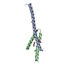
| ||||||||
|---|---|---|---|---|---|---|---|---|---|
| 1 | 
| ||||||||
| 2 | 
| ||||||||
| Unit cell |
|
-Components
| #1: Protein | Mass: 23233.215 Da / Num. of mol.: 2 Source method: isolated from a genetically manipulated source Source: (gene. exp.) Campylobacter jejuni (Campylobacter) / Gene: Cj0788 / Plasmid: CajeA.19923.a.LA1 / Production host: #2: Chemical |   Mass: 90.121 Da / Num. of mol.: 2 / Source method: obtained synthetically / Formula: C4H10O2 Mass: 90.121 Da / Num. of mol.: 2 / Source method: obtained synthetically / Formula: C4H10O2#3: Chemical | ChemComp-MN / |   Mass: 54.938 Da / Num. of mol.: 1 / Source method: obtained synthetically / Formula: Mn Mass: 54.938 Da / Num. of mol.: 1 / Source method: obtained synthetically / Formula: Mn#4: Chemical | ChemComp-CL / |   Mass: 35.453 Da / Num. of mol.: 1 / Source method: obtained synthetically / Formula: Cl Mass: 35.453 Da / Num. of mol.: 1 / Source method: obtained synthetically / Formula: Cl#5: Water | ChemComp-HOH / |  Mass: 18.015 Da / Num. of mol.: 4 / Source method: isolated from a natural source / Formula: H2O Mass: 18.015 Da / Num. of mol.: 4 / Source method: isolated from a natural source / Formula: H2OHas ligand of interest | N | |
|---|
-Experimental details
-Experiment
| Experiment | Method: X-RAY DIFFRACTION / Number of used crystals: 1 |
|---|
- Sample preparation
| Crystal grow | Temperature: 291 K / Method: vapor diffusion, sitting drop / pH: 6.5 Details: Morpheus Fusion A2: 20% v/v Ethylene glycol; 10 % w/v PEG 8000, 0.04M Imidazole; 0.06M MES monohydrate (acid), pH 6.5, 0.02M 1,6-Hexanediol; 0.02M 1-Butanol, 0.02M 1,2-Propanediol; 0.02M 2- ...Details: Morpheus Fusion A2: 20% v/v Ethylene glycol; 10 % w/v PEG 8000, 0.04M Imidazole; 0.06M MES monohydrate (acid), pH 6.5, 0.02M 1,6-Hexanediol; 0.02M 1-Butanol, 0.02M 1,2-Propanediol; 0.02M 2-Propanol; 0.02M 1,4-Butanediol; 0.02M 1,3-Propanediol, 5 mM MnCl2, 5 mM CoCl2 , 5 mM NiCl2, 5 mM Zn(OAc)2 . CajeA.19923.a.LA1.PB00122 at 27 mg/mL. Plate: 13443 well A2, drop 1. Puck: PSL-1703, Cryo: Direct. Mass spectrometry indicated a truncated fragment of the full-length protein ~7,142 Da-7,161 Da. The electron density best fit residues approximately spanning Ser 106-Lys 165. |
|---|
-Data collection
| Diffraction | Mean temperature: 100 K / Serial crystal experiment: N |
|---|---|
| Diffraction source | Source: SYNCHROTRON / Site: NSLS-II / Beamline: 19-ID / Wavelength: 0.9795 Å |
| Detector | Type: DECTRIS EIGER2 XE 9M / Detector: PIXEL / Date: Oct 9, 2023 |
| Radiation | Monochromator: Double Crystal Si 111 / Protocol: SINGLE WAVELENGTH / Monochromatic (M) / Laue (L): M / Scattering type: x-ray |
| Radiation wavelength | Wavelength: 0.9795 Å / Relative weight: 1 |
| Reflection | Resolution: 2.45→46.96 Å / Num. obs: 5660 / % possible obs: 99.9 % / Redundancy: 6.1 % / CC1/2: 0.998 / Rmerge(I) obs: 0.09 / Rpim(I) all: 0.04 / Rrim(I) all: 0.098 / Χ2: 1.01 / Net I/σ(I): 11.8 / Num. measured all: 34581 |
| Reflection shell | Resolution: 2.45→2.51 Å / % possible obs: 100 % / Redundancy: 6.3 % / Rmerge(I) obs: 0.779 / Num. measured all: 2472 / Num. unique obs: 394 / CC1/2: 0.892 / Rpim(I) all: 0.337 / Rrim(I) all: 0.851 / Χ2: 0.96 / Net I/σ(I) obs: 2.5 |
- Processing
| Software |
| |||||||||||||||||||||||||||||||||||||||||||||||||||||||||||||||||||||||||||
|---|---|---|---|---|---|---|---|---|---|---|---|---|---|---|---|---|---|---|---|---|---|---|---|---|---|---|---|---|---|---|---|---|---|---|---|---|---|---|---|---|---|---|---|---|---|---|---|---|---|---|---|---|---|---|---|---|---|---|---|---|---|---|---|---|---|---|---|---|---|---|---|---|---|---|---|---|
| Refinement | Method to determine structure: MOLECULAR REPLACEMENT / Resolution: 2.45→44.52 Å / SU ML: 0.2 / Cross valid method: FREE R-VALUE / σ(F): 1.35 / Phase error: 22.16 / Stereochemistry target values: ML
| |||||||||||||||||||||||||||||||||||||||||||||||||||||||||||||||||||||||||||
| Solvent computation | Shrinkage radii: 0.9 Å / VDW probe radii: 1.1 Å / Solvent model: FLAT BULK SOLVENT MODEL | |||||||||||||||||||||||||||||||||||||||||||||||||||||||||||||||||||||||||||
| Refinement step | Cycle: LAST / Resolution: 2.45→44.52 Å
| |||||||||||||||||||||||||||||||||||||||||||||||||||||||||||||||||||||||||||
| Refine LS restraints |
| |||||||||||||||||||||||||||||||||||||||||||||||||||||||||||||||||||||||||||
| LS refinement shell |
| |||||||||||||||||||||||||||||||||||||||||||||||||||||||||||||||||||||||||||
| Refinement TLS params. | Method: refined / Refine-ID: X-RAY DIFFRACTION
| |||||||||||||||||||||||||||||||||||||||||||||||||||||||||||||||||||||||||||
| Refinement TLS group |
|
