 Movie
Movie Controller

[English] 日本語
 Yorodumi
- PDB-8uz4: Crystal Structure of macrophage migration inhibitory factor (MIF)... -
+ Open data

- Basic information
| Entry | Database: PDB / ID: 8uz4 | |||||||||
|---|---|---|---|---|---|---|---|---|---|---|
| Title | Crystal Structure of macrophage migration inhibitory factor (MIF) from Trichomonas vaginalis (Apo, P41212 form) | |||||||||
 Components Components | MACROPHAGE MIGRATION INHIBITORY FACTOR | |||||||||
 Keywords Keywords | CYTOKINE / SSGCID / STRUCTURAL GENOMICS / SEATTLE STRUCTURAL GENOMICS CENTER FOR INFECTIOUS DISEASE / macrophage migration inhibitory factor | |||||||||
| Function / homology | phenylpyruvate tautomerase / L-dopachrome isomerase / phenylpyruvate tautomerase activity / Macrophage migration inhibitory factor / Macrophage migration inhibitory factor (MIF) / Tautomerase/MIF superfamily / cytokine activity / extracellular space / L-dopachrome isomerase Function and homology information Function and homology information | |||||||||
| Biological species |  Trichomonas vaginalis (eukaryote) Trichomonas vaginalis (eukaryote) | |||||||||
| Method |  X-RAY DIFFRACTION / SYNCHROTRON / MOLECULAR REPLACEMENT / Resolution: 2.4 Å X-RAY DIFFRACTION / SYNCHROTRON / MOLECULAR REPLACEMENT / Resolution: 2.4 Å | |||||||||
 Authors Authors | Seattle Structural Genomics Center for Infectious Disease / Seattle Structural Genomics Center for Infectious Disease (SSGCID) | |||||||||
| Funding support |  United States, 2items United States, 2items
| |||||||||
 Citation Citation | Journal: Acta Crystallogr.,Sect.F / Year: 2024 Title: Structures of Trichomonas vaginalis macrophage migratory inhibitory factor. Authors: Srivastava, A. / Nair, A. / Dawson, O.C.O. / Gao, R. / Liu, L. / Craig, J.K. / Battaile, K.P. / Harmon, E.K. / Barrett, L.K. / Van Voorhis, W.C. / Subramanian, S. / Myler, P.J. / Lovell, S. ...Authors: Srivastava, A. / Nair, A. / Dawson, O.C.O. / Gao, R. / Liu, L. / Craig, J.K. / Battaile, K.P. / Harmon, E.K. / Barrett, L.K. / Van Voorhis, W.C. / Subramanian, S. / Myler, P.J. / Lovell, S. / Asojo, O.A. / Darwiche, R. | |||||||||
| History |
|
- Structure visualization
| Structure viewer | Molecule: MolmilJmol/JSmol |
|---|
- Downloads & links
-Download
| PDBx/mmCIF format | 8uz4.cif.gz | 147 KB | Display | PDBx/mmCIF format |
|---|---|---|---|---|
| PDB format | pdb8uz4.ent.gz | 115.3 KB | Display | PDB format |
| PDBx/mmJSON format | 8uz4.json.gz | Tree view | PDBx/mmJSON format | |
| Others |  Other downloads Other downloads |
-Validation report
| Arichive directory | https://data.pdbj.org/pub/pdb/validation_reports/uz/8uz4ftp://data.pdbj.org/pub/pdb/validation_reports/uz/8uz4 | HTTPS FTP |
|---|
-Related structure data


-Links
 PDBj
PDBj- Assembly
| Deposited unit | 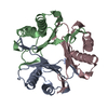
| ||||||||
|---|---|---|---|---|---|---|---|---|---|
| 1 |
| ||||||||
| Unit cell |
|
-Components
| #1: Protein | Mass: 14842.798 Da / Num. of mol.: 3 Source method: isolated from a genetically manipulated source Source: (gene. exp.) Trichomonas vaginalis (eukaryote) / Strain: G3 / Gene: TVAG_219770 / Plasmid: TrvaA.00834.a.UX1 / Production host:  #2: Water | ChemComp-HOH / |  Mass: 18.015 Da / Num. of mol.: 3 / Source method: isolated from a natural source / Formula: H2O Mass: 18.015 Da / Num. of mol.: 3 / Source method: isolated from a natural source / Formula: H2OHas protein modification | N | |
|---|
-Experimental details
-Experiment
| Experiment | Method: X-RAY DIFFRACTION / Number of used crystals: 1 |
|---|
- Sample preparation
| Crystal | Density Matthews: 2.22 Å3/Da / Density % sol: 44.49 % |
|---|---|
| Crystal grow | Temperature: 291 K / Method: vapor diffusion, sitting drop / pH: 8.5 Details: Berkeley D5: 100 mM Hepes free acid/ Sodium hydroxide pH 7.5 200 mM Ammonium acetate 25% (w/v) PEG 3350. TrvaA.00834.a.UX1.PW39224 at 35.4 mg/mL. Plate: 13617 well D5 drop 2, Puck: PSL-1907, ...Details: Berkeley D5: 100 mM Hepes free acid/ Sodium hydroxide pH 7.5 200 mM Ammonium acetate 25% (w/v) PEG 3350. TrvaA.00834.a.UX1.PW39224 at 35.4 mg/mL. Plate: 13617 well D5 drop 2, Puck: PSL-1907, Cryo: 20% PEG 200 + 80% crystallant |
-Data collection
| Diffraction | Mean temperature: 100 K / Serial crystal experiment: N |
|---|---|
| Diffraction source | Source: SYNCHROTRON / Site: NSLS-II / Beamline: 19-ID / Wavelength: 0.9795 Å |
| Detector | Type: DECTRIS EIGER2 XE 9M / Detector: PIXEL / Date: Oct 9, 2023 |
| Radiation | Monochromator: Double Crystal Si 111 / Protocol: SINGLE WAVELENGTH / Monochromatic (M) / Laue (L): M / Scattering type: x-ray |
| Radiation wavelength | Wavelength: 0.9795 Å / Relative weight: 1 |
| Reflection | Resolution: 2.4→80.71 Å / Num. obs: 16349 / % possible obs: 100 % / Redundancy: 23.2 % / CC1/2: 0.999 / Rmerge(I) obs: 0.091 / Rpim(I) all: 0.02 / Rrim(I) all: 0.093 / Χ2: 1.01 / Net I/σ(I): 21.2 / Num. measured all: 379848 |
| Reflection shell | Resolution: 2.4→2.46 Å / % possible obs: 100 % / Redundancy: 24.6 % / Rmerge(I) obs: 2.689 / Num. measured all: 29417 / Num. unique obs: 1195 / CC1/2: 0.627 / Rpim(I) all: 0.549 / Rrim(I) all: 2.745 / Χ2: 1.01 / Net I/σ(I) obs: 1.6 |
- Processing
| Software |
| ||||||||||||||||||||||||||||||||||||||||||||||||||||||||||||||||||||||||||||||||||||||||||||||||||||
|---|---|---|---|---|---|---|---|---|---|---|---|---|---|---|---|---|---|---|---|---|---|---|---|---|---|---|---|---|---|---|---|---|---|---|---|---|---|---|---|---|---|---|---|---|---|---|---|---|---|---|---|---|---|---|---|---|---|---|---|---|---|---|---|---|---|---|---|---|---|---|---|---|---|---|---|---|---|---|---|---|---|---|---|---|---|---|---|---|---|---|---|---|---|---|---|---|---|---|---|---|---|
| Refinement | Method to determine structure: MOLECULAR REPLACEMENT / Resolution: 2.4→67.17 Å / SU ML: 0.43 / Cross valid method: FREE R-VALUE / σ(F): 1.35 / Phase error: 32.68 / Stereochemistry target values: ML
| ||||||||||||||||||||||||||||||||||||||||||||||||||||||||||||||||||||||||||||||||||||||||||||||||||||
| Solvent computation | Shrinkage radii: 0.9 Å / VDW probe radii: 1.1 Å / Solvent model: FLAT BULK SOLVENT MODEL | ||||||||||||||||||||||||||||||||||||||||||||||||||||||||||||||||||||||||||||||||||||||||||||||||||||
| Refinement step | Cycle: LAST / Resolution: 2.4→67.17 Å
| ||||||||||||||||||||||||||||||||||||||||||||||||||||||||||||||||||||||||||||||||||||||||||||||||||||
| Refine LS restraints |
| ||||||||||||||||||||||||||||||||||||||||||||||||||||||||||||||||||||||||||||||||||||||||||||||||||||
| LS refinement shell |
| ||||||||||||||||||||||||||||||||||||||||||||||||||||||||||||||||||||||||||||||||||||||||||||||||||||
| Refinement TLS params. | Method: refined / Refine-ID: X-RAY DIFFRACTION
| ||||||||||||||||||||||||||||||||||||||||||||||||||||||||||||||||||||||||||||||||||||||||||||||||||||
| Refinement TLS group |
|
