 Movie
Movie Controller

[English] 日本語
 Yorodumi
- PDB-8uwv: Crystal structure of BT3984 SusD-like from Bacteroides thetaiotao... -
+ Open data

- Basic information
| Entry | Database: PDB / ID: 8uwv | ||||||
|---|---|---|---|---|---|---|---|
| Title | Crystal structure of BT3984 SusD-like from Bacteroides thetaiotaomicron VPI-5482 at 1.1 A resolution (Space group P21) | ||||||
 Components Components | BT3984 SusD-like | ||||||
 Keywords Keywords | SUGAR BINDING PROTEIN / SusD-like / High-mannose N-glycan PUL / Bacteroides thetaiotaomicron | ||||||
| Function / homology | SusD-like / Susd and RagB outer membrane lipoprotein / Prokaryotic membrane lipoprotein lipid attachment site profile. / Tetratricopeptide-like helical domain superfamily / metal ion binding / SusD homolog Function and homology information Function and homology information | ||||||
| Biological species |  Bacteroides thetaiotaomicron VPI-5482 (bacteria) Bacteroides thetaiotaomicron VPI-5482 (bacteria) | ||||||
| Method |  X-RAY DIFFRACTION / SYNCHROTRON / MOLECULAR REPLACEMENT / Resolution: 1.08 Å X-RAY DIFFRACTION / SYNCHROTRON / MOLECULAR REPLACEMENT / Resolution: 1.08 Å | ||||||
 Authors Authors | Sastre, D.E. / Navarro, M.V.A.S. / Sundberg, E.J. | ||||||
| Funding support |  United States, 1items United States, 1items
| ||||||
 Citation Citation | Journal: To Be Published Title: Crystal structure of BT3984 SusD-like from Bacteroides thetaiotaomicron VPI-5482 at 1.1 A resolution (Space group P21) Authors: Sastre, D.E. / Navarro, M.V.A.S. / Sundberg, E.J. | ||||||
| History |
|
- Structure visualization
| Structure viewer | Molecule: MolmilJmol/JSmol |
|---|
- Downloads & links
-Download
| PDBx/mmCIF format | 8uwv.cif.gz | 613.4 KB | Display | PDBx/mmCIF format |
|---|---|---|---|---|
| PDB format | pdb8uwv.ent.gz | 509.8 KB | Display | PDB format |
| PDBx/mmJSON format | 8uwv.json.gz | Tree view | PDBx/mmJSON format | |
| Others |  Other downloads Other downloads |
-Validation report
| Summary document | 8uwv_validation.pdf.gz | 1.1 MB | Display | wwPDB validaton report |
|---|---|---|---|---|
| Full document | 8uwv_full_validation.pdf.gz | 1.1 MB | Display | |
| Data in XML | 8uwv_validation.xml.gz | 53.7 KB | Display | |
| Data in CIF | 8uwv_validation.cif.gz | 84.2 KB | Display | |
| Arichive directory | https://data.pdbj.org/pub/pdb/validation_reports/uw/8uwvftp://data.pdbj.org/pub/pdb/validation_reports/uw/8uwv | HTTPS FTP |
-Related structure data
| Similar structure data |
|---|
-Links
 PDBj
PDBj


- Assembly
| Deposited unit | 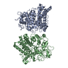
| ||||||||
|---|---|---|---|---|---|---|---|---|---|
| 1 | 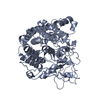
| ||||||||
| 2 | 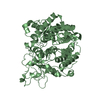
| ||||||||
| Unit cell |
|
-Components
| #1: Protein | Mass: 56809.812 Da / Num. of mol.: 2 Source method: isolated from a genetically manipulated source Source: (gene. exp.) Bacteroides thetaiotaomicron VPI-5482 (bacteria)Gene: BT_3984 / Production host: #2: Chemical | ChemComp-MG / |   Mass: 24.305 Da / Num. of mol.: 1 / Source method: obtained synthetically / Formula: Mg / Feature type: SUBJECT OF INVESTIGATION Mass: 24.305 Da / Num. of mol.: 1 / Source method: obtained synthetically / Formula: Mg / Feature type: SUBJECT OF INVESTIGATION#3: Water | ChemComp-HOH / |  Mass: 18.015 Da / Num. of mol.: 1572 / Source method: isolated from a natural source / Formula: H2O Mass: 18.015 Da / Num. of mol.: 1572 / Source method: isolated from a natural source / Formula: H2OHas ligand of interest | Y | |
|---|
-Experimental details
-Experiment
| Experiment | Method: X-RAY DIFFRACTION / Number of used crystals: 1 |
|---|
- Sample preparation
| Crystal | Density Matthews: 2.03 Å3/Da / Density % sol: 39.4 % |
|---|---|
| Crystal grow | Temperature: 291 K / Method: vapor diffusion, sitting drop Details: 0.2 M Magnesium chloride hexahydrate, 0.1 M BIS-TRIS pH 6.5, 25% w/v Polyethylene glycol 3,350 (INDEX G-11) |
-Data collection
| Diffraction | Mean temperature: 100 K / Serial crystal experiment: N |
|---|---|
| Diffraction source | Source: SYNCHROTRON / Site: LNLS SIRIUS / Beamline: MANACA / Wavelength: 0.97718 Å |
| Detector | Type: DECTRIS PILATUS 2M / Detector: PIXEL / Date: Jul 7, 2021 |
| Radiation | Protocol: SINGLE WAVELENGTH / Monochromatic (M) / Laue (L): M / Scattering type: x-ray |
| Radiation wavelength | Wavelength: 0.97718 Å / Relative weight: 1 |
| Reflection | Resolution: 1.08→46.18 Å / Num. obs: 355545 / % possible obs: 91.5 % / Redundancy: 2.5 % / Biso Wilson estimate: 9.03 Å2 / CC1/2: 0.999 / Rmerge(I) obs: 0.043 / Rrim(I) all: 0.0476 / Net I/σ(I): 19.05 |
| Reflection shell | Resolution: 1.08→1.119 Å / Redundancy: 1.3 % / Rmerge(I) obs: 0.21 / Mean I/σ(I) obs: 3.63 / Num. unique obs: 15911 / CC1/2: 0.943 / Rrim(I) all: 0.254 / % possible all: 41.19 |
- Processing
| Software |
| |||||||||||||||||||||||||||||||||||||||||||||||||||||||||||||||||||||||||||||||||||||||||||||||||||||||||||||||||||||||||||||||||||||||||||||||||||||||||||||||||||||||||||||||||||||||||||||||||||||||||||||||||||||||||
|---|---|---|---|---|---|---|---|---|---|---|---|---|---|---|---|---|---|---|---|---|---|---|---|---|---|---|---|---|---|---|---|---|---|---|---|---|---|---|---|---|---|---|---|---|---|---|---|---|---|---|---|---|---|---|---|---|---|---|---|---|---|---|---|---|---|---|---|---|---|---|---|---|---|---|---|---|---|---|---|---|---|---|---|---|---|---|---|---|---|---|---|---|---|---|---|---|---|---|---|---|---|---|---|---|---|---|---|---|---|---|---|---|---|---|---|---|---|---|---|---|---|---|---|---|---|---|---|---|---|---|---|---|---|---|---|---|---|---|---|---|---|---|---|---|---|---|---|---|---|---|---|---|---|---|---|---|---|---|---|---|---|---|---|---|---|---|---|---|---|---|---|---|---|---|---|---|---|---|---|---|---|---|---|---|---|---|---|---|---|---|---|---|---|---|---|---|---|---|---|---|---|---|---|---|---|---|---|---|---|---|---|---|---|---|---|---|---|---|
| Refinement | Method to determine structure: MOLECULAR REPLACEMENT / Resolution: 1.08→46.18 Å / SU ML: 0.06 / Cross valid method: FREE R-VALUE / σ(F): 1.39 / Phase error: 12.23 / Stereochemistry target values: ML
| |||||||||||||||||||||||||||||||||||||||||||||||||||||||||||||||||||||||||||||||||||||||||||||||||||||||||||||||||||||||||||||||||||||||||||||||||||||||||||||||||||||||||||||||||||||||||||||||||||||||||||||||||||||||||
| Solvent computation | Shrinkage radii: 0.9 Å / VDW probe radii: 1.11 Å / Solvent model: FLAT BULK SOLVENT MODEL | |||||||||||||||||||||||||||||||||||||||||||||||||||||||||||||||||||||||||||||||||||||||||||||||||||||||||||||||||||||||||||||||||||||||||||||||||||||||||||||||||||||||||||||||||||||||||||||||||||||||||||||||||||||||||
| Refinement step | Cycle: LAST / Resolution: 1.08→46.18 Å
| |||||||||||||||||||||||||||||||||||||||||||||||||||||||||||||||||||||||||||||||||||||||||||||||||||||||||||||||||||||||||||||||||||||||||||||||||||||||||||||||||||||||||||||||||||||||||||||||||||||||||||||||||||||||||
| Refine LS restraints |
| |||||||||||||||||||||||||||||||||||||||||||||||||||||||||||||||||||||||||||||||||||||||||||||||||||||||||||||||||||||||||||||||||||||||||||||||||||||||||||||||||||||||||||||||||||||||||||||||||||||||||||||||||||||||||
| LS refinement shell |
|
