 Movie
Movie Controller


+ Open data

- Basic information
| Entry | Database: PDB / ID: 8uuk | |||||||||||||||||||||||||||||||||
|---|---|---|---|---|---|---|---|---|---|---|---|---|---|---|---|---|---|---|---|---|---|---|---|---|---|---|---|---|---|---|---|---|---|---|
| Title | Pendrin in apo | |||||||||||||||||||||||||||||||||
 Components Components | Pendrin | |||||||||||||||||||||||||||||||||
 Keywords Keywords | TRANSPORT PROTEIN / chloride transport / iodide transport / bicarbonate transport | |||||||||||||||||||||||||||||||||
| Function / homology |  Function and homology information Function and homology informationiodide transmembrane transporter activity / secondary active sulfate transmembrane transporter activity / regulation of pH / chloride:bicarbonate antiporter activity / brush border membrane / regulation of protein localization / extracellular exosome Similarity search - Function | |||||||||||||||||||||||||||||||||
| Biological species |  | |||||||||||||||||||||||||||||||||
| Method | ELECTRON MICROSCOPY / single particle reconstruction / cryo EM / Resolution: 2.5 Å | |||||||||||||||||||||||||||||||||
 Authors Authors | Wang, L. / Hoang, A. / Zhou, M. | |||||||||||||||||||||||||||||||||
| Funding support |  United States, 2items United States, 2items
| |||||||||||||||||||||||||||||||||
 Citation Citation | Journal: Nat Commun / Year: 2024 Title: Mechanism of anion exchange and small-molecule inhibition of pendrin. Authors: Lie Wang / Anthony Hoang / Eva Gil-Iturbe / Arthur Laganowsky / Matthias Quick / Ming Zhou / Abstract: Pendrin (SLC26A4) is an anion exchanger that mediates bicarbonate (HCO) exchange for chloride (Cl) and is crucial for maintaining pH and salt homeostasis in the kidney, lung, and cochlea. Pendrin ...Pendrin (SLC26A4) is an anion exchanger that mediates bicarbonate (HCO) exchange for chloride (Cl) and is crucial for maintaining pH and salt homeostasis in the kidney, lung, and cochlea. Pendrin also exports iodide (I) in the thyroid gland. Pendrin mutations in humans lead to Pendred syndrome, causing hearing loss and goiter. Inhibition of pendrin is a validated approach for attenuating airway hyperresponsiveness in asthma and for treating hypertension. However, the mechanism of anion exchange and its inhibition by drugs remains poorly understood. We applied cryo-electron microscopy to determine structures of pendrin from Sus scrofa in the presence of either Cl, I, HCO or in the apo-state. The structures reveal two anion-binding sites in each protomer, and functional analyses show both sites are involved in anion exchange. The structures also show interactions between the Sulfate Transporter and Anti-Sigma factor antagonist (STAS) and transmembrane domains, and mutational studies suggest a regulatory role. We also determine the structure of pendrin in a complex with niflumic acid (NFA), which uncovers a mechanism of inhibition by competing with anion binding and impeding the structural changes necessary for anion exchange. These results reveal directions for understanding the mechanisms of anion selectivity and exchange and their regulations by the STAS domain. This work also establishes a foundation for analyzing the pathophysiology of mutations associated with Pendred syndrome. | |||||||||||||||||||||||||||||||||
| History |
|
- Structure visualization
| Structure viewer | Molecule: MolmilJmol/JSmol |
|---|
- Downloads & links
-Download
| PDBx/mmCIF format | 8uuk.cif.gz | 271.5 KB | Display | PDBx/mmCIF format |
|---|---|---|---|---|
| PDB format | pdb8uuk.ent.gz | 212.6 KB | Display | PDB format |
| PDBx/mmJSON format | 8uuk.json.gz | Tree view | PDBx/mmJSON format | |
| Others |  Other downloads Other downloads |
-Validation report
| Arichive directory | https://data.pdbj.org/pub/pdb/validation_reports/uu/8uukftp://data.pdbj.org/pub/pdb/validation_reports/uu/8uuk | HTTPS FTP |
|---|
-Related structure data
| Related structure data |  42588MC 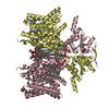 8sgwC 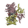 8sh3C  8shcC 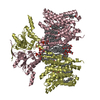 8sieC M: map data used to model this data C: citing same article ( |
|---|---|
| Similar structure data |


-Links
 PDBj
PDBj


- Assembly
| Deposited unit | 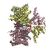
|
|---|---|
| 1 |
|
-Components
| #1: Protein | Mass: 86113.273 Da / Num. of mol.: 2 Source method: isolated from a genetically manipulated source Source: (gene. exp.)  Trichoplusia ni (cabbage looper) / References: UniProt: A0A8D0Z6H8 Trichoplusia ni (cabbage looper) / References: UniProt: A0A8D0Z6H8#2: Chemical | ChemComp-CLR /   Mass: 386.654 Da / Num. of mol.: 12 / Source method: obtained synthetically / Formula: C27H46O Mass: 386.654 Da / Num. of mol.: 12 / Source method: obtained synthetically / Formula: C27H46O#3: Chemical | ChemComp-LBN /   Mass: 760.076 Da / Num. of mol.: 14 / Source method: obtained synthetically / Formula: C42H82NO8P / Comment: phospholipid*YM Mass: 760.076 Da / Num. of mol.: 14 / Source method: obtained synthetically / Formula: C42H82NO8P / Comment: phospholipid*YM#4: Chemical | ChemComp-AV0 /   Mass: 1005.188 Da / Num. of mol.: 4 / Source method: obtained synthetically / Formula: C47H88O22 / Feature type: SUBJECT OF INVESTIGATION Mass: 1005.188 Da / Num. of mol.: 4 / Source method: obtained synthetically / Formula: C47H88O22 / Feature type: SUBJECT OF INVESTIGATIONHas ligand of interest | Y | Has protein modification | N | |
|---|
-Experimental details
-Experiment
| Experiment | Method: ELECTRON MICROSCOPY |
|---|---|
| EM experiment | Aggregation state: PARTICLE / 3D reconstruction method: single particle reconstruction |
- Sample preparation
| Component | Name: homodimer of Pendrin / Type: ORGANELLE OR CELLULAR COMPONENT / Entity ID: #1 / Source: RECOMBINANT |
|---|---|
| Molecular weight | Value: 0.075 MDa / Experimental value: NO |
| Source (natural) | Organism: |
| Source (recombinant) | Organism: Trichoplusia ni (cabbage looper) |
| Buffer solution | pH: 7.5 |
| Specimen | Conc.: 10 mg/ml / Embedding applied: NO / Shadowing applied: NO / Staining applied: NO / Vitrification applied: YES |
| Vitrification | Cryogen name: ETHANE |
- Electron microscopy imaging
| Experimental equipment |  Model: Titan Krios / Image courtesy: FEI Company |
|---|---|
| Microscopy | Model: FEI TITAN KRIOS |
| Electron gun | Electron source:  FIELD EMISSION GUN / Accelerating voltage: 300 kV / Illumination mode: FLOOD BEAM FIELD EMISSION GUN / Accelerating voltage: 300 kV / Illumination mode: FLOOD BEAM |
| Electron lens | Mode: BRIGHT FIELD / Nominal defocus max: 2000 nm / Nominal defocus min: 800 nm |
| Image recording | Electron dose: 50 e/Å2 / Film or detector model: GATAN K3 BIOQUANTUM (6k x 4k) |
- Processing
| EM software | Name: PHENIX / Category: model refinement | ||||||||||||||||||||||||
|---|---|---|---|---|---|---|---|---|---|---|---|---|---|---|---|---|---|---|---|---|---|---|---|---|---|
| CTF correction | Type: PHASE FLIPPING AND AMPLITUDE CORRECTION | ||||||||||||||||||||||||
| 3D reconstruction | Resolution: 2.5 Å / Resolution method: FSC 0.143 CUT-OFF / Num. of particles: 494226 / Symmetry type: POINT | ||||||||||||||||||||||||
| Refine LS restraints |
|
