 Movie
Movie Controller


+ Open data

- Basic information
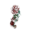
| Entry | Database: PDB / ID: 8uud | ||||||
|---|---|---|---|---|---|---|---|
| Title | BCX2627 complexed with human FVIIa and soluble Tissue Factor. | ||||||
 Components Components |
| ||||||
 Keywords Keywords | BLOOD CLOTTING / Factor VIIA & soluble Tissue factor | ||||||
| Function / homology |  Function and homology information Function and homology informationactivation of plasma proteins involved in acute inflammatory response / activation of blood coagulation via clotting cascade / coagulation factor VIIa / response to Thyroid stimulating hormone / response to astaxanthin / response to thyrotropin-releasing hormone / response to 2,3,7,8-tetrachlorodibenzodioxine / response to carbon dioxide / response to genistein / serine-type peptidase complex ...activation of plasma proteins involved in acute inflammatory response / activation of blood coagulation via clotting cascade / coagulation factor VIIa / response to Thyroid stimulating hormone / response to astaxanthin / response to thyrotropin-releasing hormone / response to 2,3,7,8-tetrachlorodibenzodioxine / response to carbon dioxide / response to genistein / serine-type peptidase complex / positive regulation of leukocyte chemotaxis / response to vitamin K / positive regulation of platelet-derived growth factor receptor signaling pathway / response to thyroxine / NGF-stimulated transcription / response to cholesterol / cytokine receptor activity / response to growth hormone / positive regulation of positive chemotaxis / Extrinsic Pathway of Fibrin Clot Formation / positive regulation of endothelial cell apoptotic process / positive regulation of blood coagulation / animal organ regeneration / positive regulation of TOR signaling / Transport of gamma-carboxylated protein precursors from the endoplasmic reticulum to the Golgi apparatus / Gamma-carboxylation of protein precursors / Removal of aminoterminal propeptides from gamma-carboxylated proteins / positive regulation of endothelial cell proliferation / serine-type peptidase activity / BMAL1:CLOCK,NPAS2 activates circadian expression / positive regulation of interleukin-8 production / circadian rhythm / protein processing / phospholipid binding / Golgi lumen / response to estrogen / cytokine-mediated signaling pathway / positive regulation of angiogenesis / blood coagulation / response to estradiol / : / protease binding / vesicle / response to hypoxia / positive regulation of cell migration / endoplasmic reticulum lumen / signaling receptor binding / external side of plasma membrane / serine-type endopeptidase activity / calcium ion binding / positive regulation of gene expression / cell surface / extracellular space / extracellular region / membrane / plasma membrane Similarity search - Function | ||||||
| Biological species |  Homo sapiens (human) Homo sapiens (human) | ||||||
| Method |  X-RAY DIFFRACTION / MOLECULAR REPLACEMENT / Resolution: 2.4 Å X-RAY DIFFRACTION / MOLECULAR REPLACEMENT / Resolution: 2.4 Å | ||||||
 Authors Authors | Krishnan, R. / Kotian, P. | ||||||
| Funding support | 1items
| ||||||
 Citation Citation | Journal: To be Published Title: Structure based Drug design of human Factor VIIa Inhibitors. Authors: Krishnan, R. | ||||||
| History |
|
- Structure visualization
| Structure viewer | Molecule: MolmilJmol/JSmol |
|---|
- Downloads & links
-Download
| PDBx/mmCIF format | 8uud.cif.gz | 144.4 KB | Display | PDBx/mmCIF format |
|---|---|---|---|---|
| PDB format | pdb8uud.ent.gz | 107.8 KB | Display | PDB format |
| PDBx/mmJSON format | 8uud.json.gz | Tree view | PDBx/mmJSON format | |
| Others |  Other downloads Other downloads |
-Validation report
| Summary document | 8uud_validation.pdf.gz | 817.6 KB | Display | wwPDB validaton report |
|---|---|---|---|---|
| Full document | 8uud_full_validation.pdf.gz | 841.9 KB | Display | |
| Data in XML | 8uud_validation.xml.gz | 31.2 KB | Display | |
| Data in CIF | 8uud_validation.cif.gz | 40.3 KB | Display | |
| Arichive directory | https://data.pdbj.org/pub/pdb/validation_reports/uu/8uudftp://data.pdbj.org/pub/pdb/validation_reports/uu/8uud | HTTPS FTP |
-Related structure data
| Related structure data | |
|---|---|
| Similar structure data |
-Links
 PDBj
PDBj


- Assembly
| Deposited unit | 
| ||||||||
|---|---|---|---|---|---|---|---|---|---|
| 1 |
| ||||||||
| Unit cell |
|
-Components
-Protein , 4 types, 4 molecules LHTU
| #1: Protein | Mass: 16359.772 Da / Num. of mol.: 1 Source method: isolated from a genetically manipulated source Source: (gene. exp.) Homo sapiens (human) / Gene: F7 / Cell line (production host): BHK / Production host:  Cricetinae (hamsters) / References: UniProt: P08709 Cricetinae (hamsters) / References: UniProt: P08709 |
|---|---|
| #2: Protein | Mass: 28103.256 Da / Num. of mol.: 1 Source method: isolated from a genetically manipulated source Source: (gene. exp.) Homo sapiens (human) / Gene: F7 / Cell line (production host): bhk / Production host: Cricetinae (hamsters) / References: UniProt: P08709, coagulation factor VIIa |
| #3: Protein | Mass: 8715.800 Da / Num. of mol.: 1 Source method: isolated from a genetically manipulated source Source: (gene. exp.) Homo sapiens (human) / Gene: F3 / Production host:  unidentified baculovirus / References: UniProt: P13726 unidentified baculovirus / References: UniProt: P13726 |
| #4: Protein | Mass: 13354.888 Da / Num. of mol.: 1 Source method: isolated from a genetically manipulated source Source: (gene. exp.) Homo sapiens (human) / Gene: F3 / Production host: unidentified baculovirus / References: UniProt: P13726 |
-Sugars , 2 types, 2 molecules 

| #5: Sugar | ChemComp-BGC /  Type: D-saccharide, beta linking / Mass: 180.156 Da / Num. of mol.: 1 Type: D-saccharide, beta linking / Mass: 180.156 Da / Num. of mol.: 1Source method: isolated from a genetically manipulated source Formula: C6H12O6 |
|---|---|
| #6: Sugar | ChemComp-FUC /  Type: L-saccharide, alpha linking / Mass: 164.156 Da / Num. of mol.: 1 Type: L-saccharide, alpha linking / Mass: 164.156 Da / Num. of mol.: 1Source method: isolated from a genetically manipulated source Formula: C6H12O5 |
-Non-polymers , 3 types, 131 molecules 

| #7: Chemical | ChemComp-CA /  Mass: 40.078 Da / Num. of mol.: 9 / Source method: obtained synthetically / Formula: Ca Mass: 40.078 Da / Num. of mol.: 9 / Source method: obtained synthetically / Formula: Ca#8: Chemical | ChemComp-XGX / ( | Mass: 484.546 Da / Num. of mol.: 1 / Source method: obtained synthetically / Formula: C28H28N4O4 #9: Water | ChemComp-HOH / | Mass: 18.015 Da / Num. of mol.: 121 / Source method: isolated from a natural source / Formula: H2O |
|---|
-Details
| Has ligand of interest | Y |
|---|---|
| Has protein modification | Y |
-Experimental details
-Experiment
| Experiment | Method: X-RAY DIFFRACTION / Number of used crystals: 1 |
|---|
- Sample preparation
| Crystal | Density Matthews: 2.66 Å3/Da / Density % sol: 53.74 % |
|---|---|
| Crystal grow | Temperature: 293 K / Method: vapor diffusion, hanging drop / pH: 6.5 Details: 14% (w/v)PEG 4K, 0.1M MgCl2, 0.1M ADA buffer, pH 6.5, VAPOR DIFFUSION, |
-Data collection
| Diffraction | Mean temperature: 98 K / Serial crystal experiment: N |
|---|---|
| Diffraction source | Source: ROTATING ANODE / Type: RIGAKU FR-D / Wavelength: 1.5418 Å |
| Detector | Type: RIGAKU RAXIS / Detector: IMAGE PLATE / Date: Jan 14, 2000 |
| Radiation | Monochromator: 1.5418 / Protocol: SINGLE WAVELENGTH / Monochromatic (M) / Laue (L): M / Scattering type: x-ray |
| Radiation wavelength | Wavelength: 1.5418 Å / Relative weight: 1 |
| Reflection | Resolution: 2.4→47.13 Å / Num. obs: 25452 / % possible obs: 90 % / Observed criterion σ(F): 3 / Redundancy: 3 % / Rmerge(I) obs: 0.1 / Net I/σ(I): 1.5 |
| Reflection shell | Resolution: 2.4→2.4 Å / Num. unique obs: 1000 / R split: 3 |
- Processing
| Software |
| ||||||||||||||||||||||||||||||||||||||||||||||||||||||||||||||||||||||||||||||||||||||||||||||||||||||||||||||||||||||||||||||||||||||||||||||||||||||||||||||||||||||||||||||||||||||
|---|---|---|---|---|---|---|---|---|---|---|---|---|---|---|---|---|---|---|---|---|---|---|---|---|---|---|---|---|---|---|---|---|---|---|---|---|---|---|---|---|---|---|---|---|---|---|---|---|---|---|---|---|---|---|---|---|---|---|---|---|---|---|---|---|---|---|---|---|---|---|---|---|---|---|---|---|---|---|---|---|---|---|---|---|---|---|---|---|---|---|---|---|---|---|---|---|---|---|---|---|---|---|---|---|---|---|---|---|---|---|---|---|---|---|---|---|---|---|---|---|---|---|---|---|---|---|---|---|---|---|---|---|---|---|---|---|---|---|---|---|---|---|---|---|---|---|---|---|---|---|---|---|---|---|---|---|---|---|---|---|---|---|---|---|---|---|---|---|---|---|---|---|---|---|---|---|---|---|---|---|---|---|---|
| Refinement | Method to determine structure: MOLECULAR REPLACEMENT / Resolution: 2.4→47.09 Å / Cor.coef. Fo:Fc: 0.931 / Cor.coef. Fo:Fc free: 0.875 / SU B: 10.301 / SU ML: 0.231 / Cross valid method: THROUGHOUT / ESU R: 0.461 / ESU R Free: 0.292 / Stereochemistry target values: MAXIMUM LIKELIHOOD / Details: HYDROGENS HAVE BEEN ADDED IN THE RIDING POSITIONS
| ||||||||||||||||||||||||||||||||||||||||||||||||||||||||||||||||||||||||||||||||||||||||||||||||||||||||||||||||||||||||||||||||||||||||||||||||||||||||||||||||||||||||||||||||||||||
| Solvent computation | Ion probe radii: 0.8 Å / Shrinkage radii: 0.8 Å / VDW probe radii: 1.2 Å / Solvent model: MASK | ||||||||||||||||||||||||||||||||||||||||||||||||||||||||||||||||||||||||||||||||||||||||||||||||||||||||||||||||||||||||||||||||||||||||||||||||||||||||||||||||||||||||||||||||||||||
| Displacement parameters | Biso mean: 36.433 Å2
| ||||||||||||||||||||||||||||||||||||||||||||||||||||||||||||||||||||||||||||||||||||||||||||||||||||||||||||||||||||||||||||||||||||||||||||||||||||||||||||||||||||||||||||||||||||||
| Refinement step | Cycle: 1 / Resolution: 2.4→47.09 Å
| ||||||||||||||||||||||||||||||||||||||||||||||||||||||||||||||||||||||||||||||||||||||||||||||||||||||||||||||||||||||||||||||||||||||||||||||||||||||||||||||||||||||||||||||||||||||
| Refine LS restraints |
|