 Movie
Movie Controller


+ Open data

- Basic information
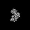
| Entry | Database: PDB / ID: 8uru | |||||||||||||||||||||||||||||||||||||||||||||
|---|---|---|---|---|---|---|---|---|---|---|---|---|---|---|---|---|---|---|---|---|---|---|---|---|---|---|---|---|---|---|---|---|---|---|---|---|---|---|---|---|---|---|---|---|---|---|
| Title | Spo11 core complex with hairpin DNA | |||||||||||||||||||||||||||||||||||||||||||||
 Components Components |
| |||||||||||||||||||||||||||||||||||||||||||||
 Keywords Keywords | DNA BINDING PROTEIN / Spo11 / Rec102 / Rec104 / Ski8 / DNA binding / Cross over. | |||||||||||||||||||||||||||||||||||||||||||||
| Function / homology |  Function and homology information Function and homology informationmeiotic DNA double-strand break processing / mRNA decay by 3' to 5' exoribonuclease / protein-DNA complex assembly / meiotic DNA double-strand break formation / Ski complex / nuclear-transcribed mRNA catabolic process, 3'-5' exonucleolytic nonsense-mediated decay / nuclear-transcribed mRNA catabolic process, non-stop decay / synaptonemal complex assembly / DNA end binding / homologous chromosome pairing at meiosis ...meiotic DNA double-strand break processing / mRNA decay by 3' to 5' exoribonuclease / protein-DNA complex assembly / meiotic DNA double-strand break formation / Ski complex / nuclear-transcribed mRNA catabolic process, 3'-5' exonucleolytic nonsense-mediated decay / nuclear-transcribed mRNA catabolic process, non-stop decay / synaptonemal complex assembly / DNA end binding / homologous chromosome pairing at meiosis / DNA topoisomerase type II (double strand cut, ATP-hydrolyzing) activity / meiosis I / reciprocal meiotic recombination / DNA topoisomerase (ATP-hydrolysing) / sporulation resulting in formation of a cellular spore / nuclear chromosome / mRNA catabolic process / nuclear-transcribed mRNA catabolic process / condensed nuclear chromosome / site of double-strand break / protein-containing complex assembly / defense response to virus / chromatin binding / DNA binding / nucleoplasm / ATP binding / metal ion binding / nucleus / cytoplasm Similarity search - Function | |||||||||||||||||||||||||||||||||||||||||||||
| Biological species |  | |||||||||||||||||||||||||||||||||||||||||||||
| Method | ELECTRON MICROSCOPY / single particle reconstruction / cryo EM / Resolution: 3.7 Å | |||||||||||||||||||||||||||||||||||||||||||||
 Authors Authors | Yu, Y. / Patel, D.J. | |||||||||||||||||||||||||||||||||||||||||||||
| Funding support |  United States, 1items United States, 1items
| |||||||||||||||||||||||||||||||||||||||||||||
 Citation Citation | Journal: Nat Struct Mol Biol / Year: 2025 Title: Cryo-EM structures of the Spo11 core complex bound to DNA. Authors: You Yu / Juncheng Wang / Kaixian Liu / Zhi Zheng / Meret Arter / Corentin Claeys Bouuaert / Stephen Pu / Dinshaw J Patel / Scott Keeney /   Abstract: DNA double-strand breaks that initiate meiotic recombination are formed by the topoisomerase-relative enzyme Spo11, supported by conserved auxiliary factors. Because high-resolution structural data ...DNA double-strand breaks that initiate meiotic recombination are formed by the topoisomerase-relative enzyme Spo11, supported by conserved auxiliary factors. Because high-resolution structural data have not been available, many questions remain about the architecture of Spo11 and its partners and how they engage with DNA. We report cryo-electron microscopy structures at up to 3.3-Å resolution of DNA-bound core complexes of Saccharomyces cerevisiae Spo11 with Rec102, Rec104 and Ski8. In these structures, monomeric core complexes make extensive contacts with the DNA backbone and with the recessed 3'-OH and first 5' overhanging nucleotide, establishing the molecular determinants of DNA end-binding specificity and providing insight into DNA cleavage preferences in vivo. The structures of individual subunits and their interfaces, supported by functional data in yeast, provide insight into the role of metal ions in DNA binding and uncover unexpected structural variation in homologs of the Top6BL component of the core complex. | |||||||||||||||||||||||||||||||||||||||||||||
| History |
|
- Structure visualization
| Structure viewer | Molecule: MolmilJmol/JSmol |
|---|
- Downloads & links
-Download
| PDBx/mmCIF format | 8uru.cif.gz | 217.2 KB | Display | PDBx/mmCIF format |
|---|---|---|---|---|
| PDB format | pdb8uru.ent.gz | 163.7 KB | Display | PDB format |
| PDBx/mmJSON format | 8uru.json.gz | Tree view | PDBx/mmJSON format | |
| Others |  Other downloads Other downloads |
-Validation report
| Arichive directory | https://data.pdbj.org/pub/pdb/validation_reports/ur/8uruftp://data.pdbj.org/pub/pdb/validation_reports/ur/8uru | HTTPS FTP |
|---|
-Related structure data
| Related structure data | 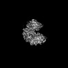 42501MC  8urqC M: map data used to model this data C: citing same article ( |
|---|---|
| Similar structure data |
-Links
 PDBj
PDBj


- Assembly
| Deposited unit | 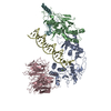
|
|---|---|
| 1 |
|
-Components
-Protein , 2 types, 2 molecules AC
| #1: Protein | Mass: 50020.109 Da / Num. of mol.: 1 Source method: isolated from a genetically manipulated source Source: (gene. exp.)   Spodoptera frugiperda (fall armyworm) / References: UniProt: P23179 Spodoptera frugiperda (fall armyworm) / References: UniProt: P23179 |
|---|---|
| #3: Protein | Mass: 44313.555 Da / Num. of mol.: 1 Source method: isolated from a genetically manipulated source Source: (gene. exp.) Spodoptera frugiperda (fall armyworm) / References: UniProt: Q02793 |
-Meiotic recombination protein ... , 2 types, 2 molecules BF
| #2: Protein | Mass: 30283.771 Da / Num. of mol.: 1 Source method: isolated from a genetically manipulated source Source: (gene. exp.) Spodoptera frugiperda (fall armyworm) / References: UniProt: Q02721 |
|---|---|
| #4: Protein | Mass: 20821.182 Da / Num. of mol.: 1 Source method: isolated from a genetically manipulated source Source: (gene. exp.) Spodoptera frugiperda (fall armyworm) / References: UniProt: P33323 |
-DNA chain / Non-polymers , 2 types, 2 molecules D
| #5: DNA chain | Mass: 14781.522 Da / Num. of mol.: 1 / Source method: obtained synthetically / Source: (synth.) |
|---|---|
| #6: Chemical | ChemComp-MG /  Mass: 24.305 Da / Num. of mol.: 1 / Source method: obtained synthetically / Formula: Mg / Feature type: SUBJECT OF INVESTIGATION Mass: 24.305 Da / Num. of mol.: 1 / Source method: obtained synthetically / Formula: Mg / Feature type: SUBJECT OF INVESTIGATION |
-Details
| Has ligand of interest | Y |
|---|---|
| Has protein modification | N |
-Experimental details
-Experiment
| Experiment | Method: ELECTRON MICROSCOPY |
|---|---|
| EM experiment | Aggregation state: PARTICLE / 3D reconstruction method: single particle reconstruction |
- Sample preparation
| Component | Name: Spo11 core complex bound to gapped DNA / Type: COMPLEX / Entity ID: #1-#5 / Source: RECOMBINANT |
|---|---|
| Molecular weight | Value: 0.14 MDa / Experimental value: NO |
| Source (natural) | Organism: |
| Source (recombinant) | Organism: Spodoptera frugiperda (fall armyworm) |
| Buffer solution | pH: 7.4 Details: 25 mM HEPES, pH 7.4, 300 mM NaCl, 5 mM EDTA, 2 mM DTT |
| Specimen | Conc.: 0.2 mg/ml / Embedding applied: NO / Shadowing applied: NO / Staining applied: NO / Vitrification applied: YES |
| Vitrification | Cryogen name: ETHANE / Humidity: 100 % / Chamber temperature: 277 K |
- Electron microscopy imaging
| Experimental equipment |  Model: Titan Krios / Image courtesy: FEI Company |
|---|---|
| Microscopy | Model: FEI TITAN KRIOS |
| Electron gun | Electron source:  FIELD EMISSION GUN / Accelerating voltage: 300 kV / Illumination mode: FLOOD BEAM FIELD EMISSION GUN / Accelerating voltage: 300 kV / Illumination mode: FLOOD BEAM |
| Electron lens | Mode: BRIGHT FIELD / Nominal defocus max: 2500 nm / Nominal defocus min: 1000 nm |
| Image recording | Average exposure time: 3 sec. / Electron dose: 53 e/Å2 / Film or detector model: GATAN K3 (6k x 4k) |
- Processing
| EM software |
| ||||||||||||||||||||||||
|---|---|---|---|---|---|---|---|---|---|---|---|---|---|---|---|---|---|---|---|---|---|---|---|---|---|
| CTF correction | Type: PHASE FLIPPING AND AMPLITUDE CORRECTION | ||||||||||||||||||||||||
| 3D reconstruction | Resolution: 3.7 Å / Resolution method: FSC 0.143 CUT-OFF / Num. of particles: 128787 / Symmetry type: POINT | ||||||||||||||||||||||||
| Refine LS restraints |
|
