 Movie
Movie Controller

[English] 日本語
 Yorodumi
- PDB-8urp: Cholinephosphotransferase in complex with CDP-choline and phospha... -
+ Open data

- Basic information
| Entry | Database: PDB / ID: 8urp | ||||||
|---|---|---|---|---|---|---|---|
| Title | Cholinephosphotransferase in complex with CDP-choline and phosphatidylcholine | ||||||
 Components Components | Cholinephosphotransferase 1 | ||||||
 Keywords Keywords | MEMBRANE PROTEIN / lipid metabolism / phospholipid synthesis / membrane protein enzyme / choline metabolism | ||||||
| Function / homology |  Function and homology information Function and homology informationSynthesis of PC / Synthesis of PE / diacylglycerol cholinephosphotransferase / diacylglycerol cholinephosphotransferase activity / CDP-choline pathway / mitochondrial outer membrane / endoplasmic reticulum membrane / endoplasmic reticulum / Golgi apparatus / metal ion binding Similarity search - Function | ||||||
| Biological species |  | ||||||
| Method | ELECTRON MICROSCOPY / single particle reconstruction / cryo EM / Resolution: 2.9 Å | ||||||
 Authors Authors | Roberts, J.R. / Maeda, S. / Ohi, M.D. | ||||||
| Funding support | 1items
| ||||||
 Citation Citation | Journal: Nat Commun / Year: 2025 Title: Structural basis for catalysis and selectivity of phospholipid synthesis by eukaryotic choline-phosphotransferase. Authors: Jacquelyn R Roberts / Yasuhiro Horibata / Frank E Kwarcinski / Vinson Lam / Ashleigh M Raczkowski / Akane Hubbard / Betsy White / Hiroyuki Sugimoto / Gregory G Tall / Melanie D Ohi / Shoji Maeda /   Abstract: Phospholipids are the most abundant component in lipid membranes and are essential for the structural and functional integrity of the cell. In eukaryotic cells, phospholipids are primarily ...Phospholipids are the most abundant component in lipid membranes and are essential for the structural and functional integrity of the cell. In eukaryotic cells, phospholipids are primarily synthesized de novo through the Kennedy pathway that involves multiple enzymatic processes. The terminal reaction is mediated by a group of cytidine-5'-diphosphate (CDP)-choline /CDP-ethanolamine-phosphotransferases (CPT/EPT) that use 1,2-diacylglycerol (DAG) and CDP-choline or CDP-ethanolamine to produce phosphatidylcholine (PC) or phosphatidylethanolamine (PE) that are the main phospholipids in eukaryotic cells. Here we present the structure of the yeast CPT1 in multiple substrate-bound states. Structural and functional analysis of these binding-sites reveal the critical residues for the DAG acyl-chain preference and the choline/ethanolamine selectivity. Additionally, we present the structure in complex with a potent inhibitor characterized in this study. The ensemble of structures allows us to propose the reaction mechanism for phospholipid biosynthesis by the family of CDP-alcohol phosphotransferases (CDP-APs). | ||||||
| History |
|
- Structure visualization
| Structure viewer | Molecule: MolmilJmol/JSmol |
|---|
- Downloads & links
-Download
| PDBx/mmCIF format | 8urp.cif.gz | 179.2 KB | Display | PDBx/mmCIF format |
|---|---|---|---|---|
| PDB format | pdb8urp.ent.gz | 140.5 KB | Display | PDB format |
| PDBx/mmJSON format | 8urp.json.gz | Tree view | PDBx/mmJSON format | |
| Others |  Other downloads Other downloads |
-Validation report
| Summary document | 8urp_validation.pdf.gz | 1.5 MB | Display | wwPDB validaton report |
|---|---|---|---|---|
| Full document | 8urp_full_validation.pdf.gz | 1.5 MB | Display | |
| Data in XML | 8urp_validation.xml.gz | 43.5 KB | Display | |
| Data in CIF | 8urp_validation.cif.gz | 59.9 KB | Display | |
| Arichive directory | https://data.pdbj.org/pub/pdb/validation_reports/ur/8urpftp://data.pdbj.org/pub/pdb/validation_reports/ur/8urp | HTTPS FTP |
-Related structure data
| Related structure data |  42496MC  8ul9C  8urtC C: citing same article ( M: map data used to model this data |
|---|---|
| Similar structure data |


-Links
 PDBj
PDBj
- Assembly
| Deposited unit | 
|
|---|---|
| 1 |
|
-Components
-Protein , 1 types, 2 molecules AB
| #1: Protein | Mass: 44147.277 Da / Num. of mol.: 2 Source method: isolated from a genetically manipulated source Source: (gene. exp.) Gene: CPT1 / Production host:   Spodoptera frugiperda (fall armyworm) / References: UniProt: P17898 Spodoptera frugiperda (fall armyworm) / References: UniProt: P17898 |
|---|
-Non-polymers , 5 types, 23 molecules 

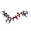
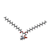

| #2: Chemical | ChemComp-MG /  Mass: 24.305 Da / Num. of mol.: 4 / Source method: obtained synthetically / Formula: Mg Mass: 24.305 Da / Num. of mol.: 4 / Source method: obtained synthetically / Formula: Mg#3: Chemical | ChemComp-PCW /  Mass: 787.121 Da / Num. of mol.: 6 / Source method: obtained synthetically / Formula: C44H85NO8P / Comment: DOPC, phospholipid*YM Mass: 787.121 Da / Num. of mol.: 6 / Source method: obtained synthetically / Formula: C44H85NO8P / Comment: DOPC, phospholipid*YM#4: Chemical |  Mass: 488.324 Da / Num. of mol.: 2 / Source method: obtained synthetically / Formula: C14H26N4O11P2 / Feature type: SUBJECT OF INVESTIGATION Mass: 488.324 Da / Num. of mol.: 2 / Source method: obtained synthetically / Formula: C14H26N4O11P2 / Feature type: SUBJECT OF INVESTIGATION#5: Chemical |  Mass: 734.039 Da / Num. of mol.: 2 / Source method: isolated from a natural source / Formula: C40H80NO8P Mass: 734.039 Da / Num. of mol.: 2 / Source method: isolated from a natural source / Formula: C40H80NO8P#6: Water | ChemComp-HOH / | Mass: 18.015 Da / Num. of mol.: 9 / Source method: isolated from a natural source / Formula: H2O |
|---|
-Details
| Has ligand of interest | Y |
|---|---|
| Has protein modification | Y |
-Experimental details
-Experiment
| Experiment | Method: ELECTRON MICROSCOPY |
|---|---|
| EM experiment | Aggregation state: PARTICLE / 3D reconstruction method: single particle reconstruction |
- Sample preparation
| Component |
| ||||||||||||||||||||||||
|---|---|---|---|---|---|---|---|---|---|---|---|---|---|---|---|---|---|---|---|---|---|---|---|---|---|
| Molecular weight |
| ||||||||||||||||||||||||
| Source (natural) |
| ||||||||||||||||||||||||
| Source (recombinant) |
| ||||||||||||||||||||||||
| Buffer solution | pH: 7.5 | ||||||||||||||||||||||||
| Specimen | Conc.: 1 mg/ml / Embedding applied: NO / Shadowing applied: NO / Staining applied: NO / Vitrification applied: YES | ||||||||||||||||||||||||
| Vitrification | Instrument: FEI VITROBOT MARK IV / Cryogen name: ETHANE / Humidity: 100 % / Chamber temperature: 278 K |


- Electron microscopy imaging
| Experimental equipment |  Model: Titan Krios / Image courtesy: FEI Company |
|---|---|
| Microscopy | Model: FEI TITAN KRIOS |
| Electron gun | Electron source:  FIELD EMISSION GUN / Accelerating voltage: 300 kV / Illumination mode: FLOOD BEAM FIELD EMISSION GUN / Accelerating voltage: 300 kV / Illumination mode: FLOOD BEAM |
| Electron lens | Mode: BRIGHT FIELD / Nominal magnification: 81000 X / Nominal defocus max: 2400 nm / Nominal defocus min: 1500 nm / Cs: 2.7 mm / Alignment procedure: COMA FREE |
| Specimen holder | Cryogen: NITROGEN / Specimen holder model: FEI TITAN KRIOS AUTOGRID HOLDER |
| Image recording | Average exposure time: 4.4 sec. / Electron dose: 60 e/Å2 / Film or detector model: GATAN K3 (6k x 4k) / Num. of grids imaged: 1 / Num. of real images: 2990 |
| EM imaging optics | Energyfilter name: GIF Bioquantum / Energyfilter slit width: 20 eV |
- Processing
| EM software |
| |||||||||||||||||||||||||||||||||||
|---|---|---|---|---|---|---|---|---|---|---|---|---|---|---|---|---|---|---|---|---|---|---|---|---|---|---|---|---|---|---|---|---|---|---|---|---|
| CTF correction | Details: Patch CTF correction / Type: PHASE FLIPPING AND AMPLITUDE CORRECTION | |||||||||||||||||||||||||||||||||||
| 3D reconstruction | Resolution: 2.9 Å / Resolution method: FSC 0.143 CUT-OFF / Num. of particles: 971309 / Symmetry type: POINT | |||||||||||||||||||||||||||||||||||
| Atomic model building | Protocol: FLEXIBLE FIT / Space: REAL / Target criteria: Cross-correlation coefficient Details: Initial model fitting was done using a model superposition function into a 3D volume in Chimera and then Phenix.real_space_refine was used for flexible fitting. | |||||||||||||||||||||||||||||||||||
| Atomic model building | Accession code: P17898 / Source name: AlphaFold / Type: in silico model | |||||||||||||||||||||||||||||||||||
| Refine LS restraints |
|
