 Movie
Movie Controller


+ Open data

- Basic information
| Entry | Database: PDB / ID: 8uqn | |||||||||
|---|---|---|---|---|---|---|---|---|---|---|
| Title | PLCb3-Gaq complex on membranes | |||||||||
 Components Components |
| |||||||||
 Keywords Keywords | SIGNALING PROTEIN / PLCb3 / Gaq | |||||||||
| Function / homology |  Function and homology information Function and homology informationphosphatidylinositol phospholipase C activity / phosphoinositide phospholipase C / Fatty Acids bound to GPR40 (FFAR1) regulate insulin secretion / Acetylcholine regulates insulin secretion / sensory perception of itch / phospholipase C-activating G protein-coupled glutamate receptor signaling pathway / phosphatidylinositol metabolic process / regulation of systemic arterial blood pressure / PLC beta mediated events / phospholipase C-activating serotonin receptor signaling pathway ...phosphatidylinositol phospholipase C activity / phosphoinositide phospholipase C / Fatty Acids bound to GPR40 (FFAR1) regulate insulin secretion / Acetylcholine regulates insulin secretion / sensory perception of itch / phospholipase C-activating G protein-coupled glutamate receptor signaling pathway / phosphatidylinositol metabolic process / regulation of systemic arterial blood pressure / PLC beta mediated events / phospholipase C-activating serotonin receptor signaling pathway / phosphatidylinositol-4,5-bisphosphate phospholipase C activity / regulation of platelet activation / entrainment of circadian clock / C-type glycerophospholipase activity / regulation of canonical Wnt signaling pathway / glutamate receptor signaling pathway / mast cell degranulation / Synthesis of IP3 and IP4 in the cytosol / phosphatidylinositol-mediated signaling / phototransduction, visible light / photoreceptor outer segment / postsynaptic cytosol / lipid catabolic process / cellular response to acidic pH / release of sequestered calcium ion into cytosol / hormone-mediated signaling pathway / molecular function activator activity / Turbulent (oscillatory, disturbed) flow shear stress activates signaling by PIEZO1 and integrins in endothelial cells / GTPase activator activity / neuropeptide signaling pathway / response to prostaglandin E / G protein-coupled receptor binding / G-protein beta/gamma-subunit complex binding / blood coagulation / G protein-coupled acetylcholine receptor signaling pathway / G beta:gamma signalling through PLC beta / Presynaptic function of Kainate receptors / Thromboxane signalling through TP receptor / G-protein activation / ADP signalling through P2Y purinoceptor 1 / Cooperation of PDCL (PhLP1) and TRiC/CCT in G-protein beta folding / heterotrimeric G-protein complex / Thrombin signalling through proteinase activated receptors (PARs) / adenylate cyclase-activating G protein-coupled receptor signaling pathway / G protein activity / Ca2+ pathway / nuclear membrane / High laminar flow shear stress activates signaling by PIEZO1 and PECAM1:CDH5:KDR in endothelial cells / molecular adaptor activity / phospholipase C-activating G protein-coupled receptor signaling pathway / G alpha (q) signalling events / Hydrolases; Acting on acid anhydrides; Acting on GTP to facilitate cellular and subcellular movement / calmodulin binding / protein stabilization / cadherin binding / G protein-coupled receptor signaling pathway / lysosomal membrane / GTPase activity / calcium ion binding / GTP binding / Golgi apparatus / protein-containing complex / extracellular exosome / membrane / metal ion binding / nucleus / plasma membrane / cytoplasm / cytosol Similarity search - Function | |||||||||
| Biological species |  Homo sapiens (human) Homo sapiens (human) | |||||||||
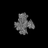
| Method | ELECTRON MICROSCOPY / single particle reconstruction / cryo EM / Resolution: 3.4 Å | |||||||||
 Authors Authors | Falzone, M.E. / MacKinnon, R. | |||||||||
| Funding support |  United States, 2items United States, 2items
| |||||||||
 Citation Citation | Journal: Proc Natl Acad Sci U S A / Year: 2023 Title: The mechanism of regulation of -catalyzed hydrolysis. Authors: Maria E Falzone / Roderick MacKinnon / Abstract: () enzymes cleave phosphatidylinositol 4,5-bisphosphate ( producing and (diacylglycerol). modulates the function of many ion channels, while and regulate intracellular Ca levels and protein ... () enzymes cleave phosphatidylinositol 4,5-bisphosphate ( producing and (diacylglycerol). modulates the function of many ion channels, while and regulate intracellular Ca levels and protein phosphorylation by protein kinase C, respectively. enzymes are under the control of G protein coupled receptor signaling through direct interactions with G proteins and and have been shown to be coincidence detectors for dual stimulation of and -coupled receptors. are aqueous-soluble cytoplasmic enzymes but partition onto the membrane surface to access their lipid substrate, complicating their functional and structural characterization. Using newly developed methods, we recently showed that activates by recruiting it to the membrane. Using these same methods, here we show that increases the catalytic rate constant, , of . Since stimulation of by depends on an autoinhibitory element (the X-Y linker), we propose that produces partial relief of the X-Y linker autoinhibition through an allosteric mechanism. We also determined membrane-bound structures of the and complexes, which show that these G proteins can bind simultaneously and independently of each other to regulate activity. The structures rationalize a finding in the enzyme assay, that costimulation by both G proteins follows a product rule of each independent stimulus. We conclude that baseline activity of is strongly suppressed, but the effect of G proteins, especially acting together, provides a robust stimulus upon G protein stimulation. | |||||||||
| History |
|
- Structure visualization
| Structure viewer | Molecule: MolmilJmol/JSmol |
|---|
- Downloads & links
-Download
| PDBx/mmCIF format | 8uqn.cif.gz | 211.2 KB | Display | PDBx/mmCIF format |
|---|---|---|---|---|
| PDB format | pdb8uqn.ent.gz | 159.3 KB | Display | PDB format |
| PDBx/mmJSON format | 8uqn.json.gz | Tree view | PDBx/mmJSON format | |
| Others |  Other downloads Other downloads |
-Validation report
| Arichive directory | https://data.pdbj.org/pub/pdb/validation_reports/uq/8uqnftp://data.pdbj.org/pub/pdb/validation_reports/uq/8uqn | HTTPS FTP |
|---|
-Related structure data
| Related structure data | 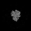 42475MC 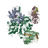 8uqoC M: map data used to model this data C: citing same article ( |
|---|---|
| Similar structure data |
-Links
 PDBj
PDBj


- Assembly
| Deposited unit | 
|
|---|---|
| 1 |
|
-Components
-Protein , 2 types, 2 molecules AB
| #1: Protein | Mass: 41676.387 Da / Num. of mol.: 1 Source method: isolated from a genetically manipulated source Source: (gene. exp.) Homo sapiens (human) / Gene: GNAQ / Production host:  Trichoplusia ni (cabbage looper) / References: UniProt: P50148 Trichoplusia ni (cabbage looper) / References: UniProt: P50148 |
|---|---|
| #2: Protein | Mass: 139104.719 Da / Num. of mol.: 1 Source method: isolated from a genetically manipulated source Source: (gene. exp.) Homo sapiens (human) / Gene: PLCB3 / Production host: Trichoplusia ni (cabbage looper)References: UniProt: Q01970, phosphoinositide phospholipase C |
-Non-polymers , 5 types, 5 molecules 


| #3: Chemical | ChemComp-GDP /  Type: RNA linking / Mass: 443.201 Da / Num. of mol.: 1 / Source method: isolated from a natural source / Formula: C10H15N5O11P2 / Comment: GDP, energy-carrying molecule*YM Type: RNA linking / Mass: 443.201 Da / Num. of mol.: 1 / Source method: isolated from a natural source / Formula: C10H15N5O11P2 / Comment: GDP, energy-carrying molecule*YM |
|---|---|
| #4: Chemical | ChemComp-ALF /  Mass: 102.975 Da / Num. of mol.: 1 / Source method: obtained synthetically / Formula: AlF4 Mass: 102.975 Da / Num. of mol.: 1 / Source method: obtained synthetically / Formula: AlF4 |
| #5: Chemical | ChemComp-MG /  Mass: 24.305 Da / Num. of mol.: 1 / Source method: obtained synthetically / Formula: Mg Mass: 24.305 Da / Num. of mol.: 1 / Source method: obtained synthetically / Formula: Mg |
| #6: Chemical | ChemComp-CA /  Mass: 40.078 Da / Num. of mol.: 1 / Source method: obtained synthetically / Formula: Ca Mass: 40.078 Da / Num. of mol.: 1 / Source method: obtained synthetically / Formula: Ca |
| #7: Water | ChemComp-HOH / Mass: 18.015 Da / Num. of mol.: 1 / Source method: isolated from a natural source / Formula: H2O |
-Details
| Has ligand of interest | N |
|---|
-Experimental details
-Experiment
| Experiment | Method: ELECTRON MICROSCOPY |
|---|---|
| EM experiment | Aggregation state: PARTICLE / 3D reconstruction method: single particle reconstruction |
- Sample preparation
| Component | Name: PLCb3-Gaq complex / Type: COMPLEX / Entity ID: #1-#2 / Source: RECOMBINANT |
|---|---|
| Molecular weight | Experimental value: NO |
| Source (natural) | Organism: Homo sapiens (human) |
| Source (recombinant) | Organism: Trichoplusia ni (cabbage looper) |
| Buffer solution | pH: 7.4 |
| Specimen | Embedding applied: NO / Shadowing applied: NO / Staining applied: NO / Vitrification applied: YES |
| Vitrification | Cryogen name: ETHANE |
- Electron microscopy imaging
| Experimental equipment |  Model: Titan Krios / Image courtesy: FEI Company |
|---|---|
| Microscopy | Model: TFS KRIOS |
| Electron gun | Electron source:  FIELD EMISSION GUN / Accelerating voltage: 300 kV / Illumination mode: FLOOD BEAM FIELD EMISSION GUN / Accelerating voltage: 300 kV / Illumination mode: FLOOD BEAM |
| Electron lens | Mode: BRIGHT FIELD / Nominal defocus max: 2500 nm / Nominal defocus min: 1500 nm |
| Image recording | Electron dose: 60 e/Å2 / Film or detector model: GATAN K3 (6k x 4k) |
- Processing
| CTF correction | Type: PHASE FLIPPING AND AMPLITUDE CORRECTION |
|---|---|
| 3D reconstruction | Resolution: 3.4 Å / Resolution method: FSC 0.143 CUT-OFF / Num. of particles: 67545 / Symmetry type: POINT |
