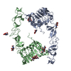
Entry Database : PDB / ID : 8ukxTitle Crystal structure the extracellular region of the epidermal growth factor receptor variant III (EGFRvIII) at pH 7.0 Epidermal growth factor receptor Keywords / / / / / Function / homology Function Domain/homology Component
/ / / / / / / / / / / / / / / / / / / / / / / / / / / / / / / / / / / / / / / / / / / / / / / / / / / / / / / / / / / / / / / / / / / / / / / / / / / / / / / / / / / / / / / / / / / / / / / / / / / / / / / / / / / / / / / / / / / / / / / / / / / / / / / / / / / / / / / / / / / / / Biological species Homo sapiens (human)Method / / / Resolution : 3.301 Å Authors Stayrook, S.E. / Ferguson, K.M. Funding support Organization Grant number Country National Institutes of Health/National Cancer Institute (NIH/NCI) R01CA198164 National Institutes of Health/National Cancer Institute (NIH/NCI) R01CA112552
Journal : Structure / Year : 2024Title : Structural insights into the role and targeting of EGFRvIII.Authors : Bagchi, A. / Stayrook, S.E. / Xenaki, K.T. / Starbird, C.A. / Doulkeridou, S. / El Khoulati, R. / Roovers, R.C. / Schmitz, K.R. / van Bergen En Henegouwen, P.M.P. / Ferguson, K.M. History Deposition Oct 15, 2023 Deposition site / Processing site Revision 1.0 Jun 12, 2024 Provider / Type Revision 1.1 Jul 3, 2024 Group / Category / citation_authorItem _citation.pdbx_database_id_DOI / _citation.pdbx_database_id_PubMed ... _citation.pdbx_database_id_DOI / _citation.pdbx_database_id_PubMed / _citation.title / _citation_author.identifier_ORCID / _citation_author.name Revision 1.2 Sep 18, 2024 Group / Category Item / _citation.page_first / _citation.page_lastRevision 1.3 Sep 25, 2024 Group / Category / Item Revision 1.4 Oct 9, 2024 Group / Category / pdbx_modification_feature / Item
Show all Show less
 Movie
Movie Controller

 Yorodumi
 Open data
 Basic information
 Components
 Keywords
 Function and homology information
 Homo sapiens (human)
 X-RAY DIFFRACTION /
  Authors
 United States, 2items
  Citation
 Structure visualization
 Downloads & links
 Other downloads
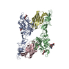
 PDBj
PDBj


 Assembly

 Spodoptera frugiperda (fall armyworm)
 Type: D-saccharide, beta linking / Mass: 221.208 Da / Num. of mol.: 2 / Source method: obtained synthetically / Formula: C8H15NO6
 Sample preparation
 Processing