 Movie
Movie Controller

[English] 日本語
 Yorodumi
- PDB-8ujl: Crystal structure of human CTDNEP1-NEP1R1 protein phosphatase complex -
+ Open data

- Basic information
| Entry | Database: PDB / ID: 8ujl | ||||||
|---|---|---|---|---|---|---|---|
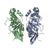
| Title | Crystal structure of human CTDNEP1-NEP1R1 protein phosphatase complex | ||||||
 Components Components | CTD nuclear envelope phosphatase 1,Nuclear envelope phosphatase-regulatory subunit 1 | ||||||
 Keywords Keywords | HYDROLASE / phosphatase / nuclear envelope | ||||||
| Function / homology |  Function and homology information Function and homology informationNem1-Spo7 phosphatase complex / Depolymerization of the Nuclear Lamina / nuclear envelope organization / positive regulation of triglyceride biosynthetic process / mitotic nuclear membrane disassembly / gamete generation / protein-serine/threonine phosphatase / protein serine/threonine phosphatase activity / mesoderm development / phosphoprotein phosphatase activity ...Nem1-Spo7 phosphatase complex / Depolymerization of the Nuclear Lamina / nuclear envelope organization / positive regulation of triglyceride biosynthetic process / mitotic nuclear membrane disassembly / gamete generation / protein-serine/threonine phosphatase / protein serine/threonine phosphatase activity / mesoderm development / phosphoprotein phosphatase activity / protein localization to nucleus / canonical Wnt signaling pathway / protein dephosphorylation / lipid droplet / lipid metabolic process / nuclear envelope / positive regulation of canonical Wnt signaling pathway / nuclear membrane / endoplasmic reticulum membrane / cytoplasm / cytosol Similarity search - Function | ||||||
| Biological species |  Homo sapiens (human) Homo sapiens (human) | ||||||
| Method |  X-RAY DIFFRACTION / SYNCHROTRON / MOLECULAR REPLACEMENT / Resolution: 1.91 Å X-RAY DIFFRACTION / SYNCHROTRON / MOLECULAR REPLACEMENT / Resolution: 1.91 Å | ||||||
 Authors Authors | Gao, S. / Airola, M.V. | ||||||
| Funding support |  United States, 1items United States, 1items
| ||||||
 Citation Citation | Journal: Proc.Natl.Acad.Sci.USA / Year: 2024 Title: Structure and mechanism of the human CTDNEP1-NEP1R1 membrane protein phosphatase complex necessary to maintain ER membrane morphology. Authors: Gao, S. / Carrasquillo Rodriguez, J.W. / Bahmanyar, S. / Airola, M.V. | ||||||
| History |
|
- Structure visualization
| Structure viewer | Molecule: MolmilJmol/JSmol |
|---|
- Downloads & links
-Download
| PDBx/mmCIF format | 8ujl.cif.gz | 176.6 KB | Display | PDBx/mmCIF format |
|---|---|---|---|---|
| PDB format | pdb8ujl.ent.gz | 117.4 KB | Display | PDB format |
| PDBx/mmJSON format | 8ujl.json.gz | Tree view | PDBx/mmJSON format | |
| Others |  Other downloads Other downloads |
-Validation report
| Arichive directory | https://data.pdbj.org/pub/pdb/validation_reports/uj/8ujlftp://data.pdbj.org/pub/pdb/validation_reports/uj/8ujl | HTTPS FTP |
|---|
-Related structure data
-Links
 PDBj
PDBj- Assembly
| Deposited unit | 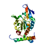
| ||||||||||||
|---|---|---|---|---|---|---|---|---|---|---|---|---|---|
| 1 |
| ||||||||||||
| Unit cell |
|
-Components
| #1: Protein | Mass: 31697.434 Da / Num. of mol.: 1 Source method: isolated from a genetically manipulated source Details: Fusion of human CTD nuclear envelope phosphatase 1 and human nuclear envelope phosphatase-regulatory subunit 1,Fusion of human CTD nuclear envelope phosphatase 1 and human nuclear envelope ...Details: Fusion of human CTD nuclear envelope phosphatase 1 and human nuclear envelope phosphatase-regulatory subunit 1,Fusion of human CTD nuclear envelope phosphatase 1 and human nuclear envelope phosphatase-regulatory subunit 1 Source: (gene. exp.) Homo sapiens (human) / Gene: CTDNEP1, CNEP1R1 / Production host:  |
|---|---|
| #2: Water | ChemComp-HOH /  Mass: 18.015 Da / Num. of mol.: 179 / Source method: isolated from a natural source / Formula: H2O Mass: 18.015 Da / Num. of mol.: 179 / Source method: isolated from a natural source / Formula: H2O |
-Experimental details
-Experiment
| Experiment | Method: X-RAY DIFFRACTION / Number of used crystals: 1 |
|---|
- Sample preparation
| Crystal | Density Matthews: 2.34 Å3/Da / Density % sol: 47.37 % |
|---|---|
| Crystal grow | Temperature: 298 K / Method: vapor diffusion, hanging drop / Details: 6% PEG 3350, 0.2 M lithium citrate, 0.4% CHAPS |
-Data collection
| Diffraction | Mean temperature: 100 K / Serial crystal experiment: N |
|---|---|
| Diffraction source | Source: SYNCHROTRON / Site: NSLS-II / Beamline: 17-ID-1 / Wavelength: 0.9201 Å |
| Detector | Type: DECTRIS EIGER X 9M / Detector: PIXEL / Date: Jul 26, 2023 / Details: KB bimorph mirrors |
| Radiation | Protocol: SINGLE WAVELENGTH / Monochromatic (M) / Laue (L): M / Scattering type: x-ray |
| Radiation wavelength | Wavelength: 0.9201 Å / Relative weight: 1 |
| Reflection | Resolution: 1.908→29.08 Å / Num. obs: 23447 / % possible obs: 99.74 % / Redundancy: 2 % / Biso Wilson estimate: 32.92 Å2 / CC1/2: 0.998 / CC star: 1 / Rmerge(I) obs: 0.05073 / Rpim(I) all: 0.05073 / Rrim(I) all: 0.07175 / Net I/σ(I): 7.92 |
| Reflection shell | Resolution: 1.908→1.976 Å / Redundancy: 2 % / Rmerge(I) obs: 0.7953 / Mean I/σ(I) obs: 0.83 / Num. unique obs: 2260 / CC1/2: 0.412 / CC star: 0.764 / Rpim(I) all: 0.7953 / Rrim(I) all: 1.125 / % possible all: 98.01 |
- Processing
| Software |
| |||||||||||||||||||||||||||||||||||||||||||||||||||||||||||||||
|---|---|---|---|---|---|---|---|---|---|---|---|---|---|---|---|---|---|---|---|---|---|---|---|---|---|---|---|---|---|---|---|---|---|---|---|---|---|---|---|---|---|---|---|---|---|---|---|---|---|---|---|---|---|---|---|---|---|---|---|---|---|---|---|---|
| Refinement | Method to determine structure: MOLECULAR REPLACEMENT / Resolution: 1.91→29.08 Å / SU ML: 0.2646 / Cross valid method: FREE R-VALUE / σ(F): 1.35 / Phase error: 20.4717 Stereochemistry target values: GeoStd + Monomer Library + CDL v1.2
| |||||||||||||||||||||||||||||||||||||||||||||||||||||||||||||||
| Solvent computation | Shrinkage radii: 0.9 Å / VDW probe radii: 1.1 Å / Solvent model: FLAT BULK SOLVENT MODEL | |||||||||||||||||||||||||||||||||||||||||||||||||||||||||||||||
| Displacement parameters | Biso mean: 44.79 Å2 | |||||||||||||||||||||||||||||||||||||||||||||||||||||||||||||||
| Refinement step | Cycle: LAST / Resolution: 1.91→29.08 Å
| |||||||||||||||||||||||||||||||||||||||||||||||||||||||||||||||
| Refine LS restraints |
| |||||||||||||||||||||||||||||||||||||||||||||||||||||||||||||||
| LS refinement shell |
| |||||||||||||||||||||||||||||||||||||||||||||||||||||||||||||||
| Refinement TLS params. | Method: refined / Origin x: -30.1150173489 Å / Origin y: -13.3384256528 Å / Origin z: -5.92299529952 Å
| |||||||||||||||||||||||||||||||||||||||||||||||||||||||||||||||
| Refinement TLS group | Selection details: all |
