 Movie
Movie Controller


+ Open data

- Basic information
| Entry | Database: PDB / ID: 8uiq | ||||||
|---|---|---|---|---|---|---|---|
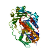
| Title | H47Q NicC with 2-mercaptopyridine ligand | ||||||
 Components Components | 6-hydroxynicotinate 3-monooxygenase | ||||||
 Keywords Keywords | OXIDOREDUCTASE / ligand bound | ||||||
| Function / homology |  Function and homology information Function and homology information6-hydroxynicotinate 3-monooxygenase / 6-hydroxynicotinate 3-monooxygenase activity / nicotinate catabolic process / FAD binding / monooxygenase activity Similarity search - Function | ||||||
| Biological species |  Pseudomonas putida KT2440 (bacteria) Pseudomonas putida KT2440 (bacteria) | ||||||
| Method |  X-RAY DIFFRACTION / SYNCHROTRON / MOLECULAR REPLACEMENT / Resolution: 2.17 Å X-RAY DIFFRACTION / SYNCHROTRON / MOLECULAR REPLACEMENT / Resolution: 2.17 Å | ||||||
 Authors Authors | Hicks, K.A. / Perry, K. / Turlington, Z.R. / Vaz Ferreira de Macedo, S. | ||||||
| Funding support |  United States, 1items United States, 1items
| ||||||
 Citation Citation | Journal: Arch.Biochem.Biophys. / Year: 2024 Title: Ligand bound structure of a 6-hydroxynicotinic acid 3-monooxygenase provides mechanistic insights. Authors: Turlington, Z.R. / Vaz Ferreira de Macedo, S. / Perry, K. / Belsky, S.L. / Faust, J.A. / Snider, M.J. / Hicks, K.A. | ||||||
| History |
|
- Structure visualization
| Structure viewer | Molecule: MolmilJmol/JSmol |
|---|
- Downloads & links
-Download
| PDBx/mmCIF format | 8uiq.cif.gz | 109.9 KB | Display | PDBx/mmCIF format |
|---|---|---|---|---|
| PDB format | pdb8uiq.ent.gz | 65.4 KB | Display | PDB format |
| PDBx/mmJSON format | 8uiq.json.gz | Tree view | PDBx/mmJSON format | |
| Others |  Other downloads Other downloads |
-Validation report
| Arichive directory | https://data.pdbj.org/pub/pdb/validation_reports/ui/8uiqftp://data.pdbj.org/pub/pdb/validation_reports/ui/8uiq | HTTPS FTP |
|---|
-Related structure data
-Links
 PDBj
PDBj- Assembly
| Deposited unit | 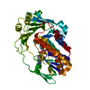
| ||||||||||||
|---|---|---|---|---|---|---|---|---|---|---|---|---|---|
| 1 |
| ||||||||||||
| Unit cell |
|
-Components
| #1: Protein | Mass: 45420.066 Da / Num. of mol.: 1 / Mutation: H47Q Source method: isolated from a genetically manipulated source Source: (gene. exp.) Pseudomonas putida KT2440 (bacteria) / Gene: nicC / Production host: |
|---|---|
| #2: Chemical | ChemComp-FAD /   Mass: 785.550 Da / Num. of mol.: 1 / Source method: obtained synthetically / Formula: C27H33N9O15P2 / Feature type: SUBJECT OF INVESTIGATION / Comment: FAD*YM Mass: 785.550 Da / Num. of mol.: 1 / Source method: obtained synthetically / Formula: C27H33N9O15P2 / Feature type: SUBJECT OF INVESTIGATION / Comment: FAD*YM |
| #3: Chemical | ChemComp-PYS /   Mass: 111.165 Da / Num. of mol.: 1 / Source method: obtained synthetically / Formula: C5H5NS / Feature type: SUBJECT OF INVESTIGATION Mass: 111.165 Da / Num. of mol.: 1 / Source method: obtained synthetically / Formula: C5H5NS / Feature type: SUBJECT OF INVESTIGATION |
| #4: Water | ChemComp-HOH /  Mass: 18.015 Da / Num. of mol.: 115 / Source method: isolated from a natural source / Formula: H2O Mass: 18.015 Da / Num. of mol.: 115 / Source method: isolated from a natural source / Formula: H2O |
| Has ligand of interest | Y |
-Experimental details
-Experiment
| Experiment | Method: X-RAY DIFFRACTION / Number of used crystals: 1 |
|---|
- Sample preparation
| Crystal | Density Matthews: 2.08 Å3/Da / Density % sol: 40.89 % |
|---|---|
| Crystal grow | Temperature: 291 K / Method: vapor diffusion, hanging drop / pH: 7.5 Details: 0.1 M Hepes, pH 7.5, 15-25% polyethylene glycol (PEG) 3350, and 0.2 M magnesium chloride hexahydrate |
-Data collection
| Diffraction |
| ||||||||||||||||||||||||
|---|---|---|---|---|---|---|---|---|---|---|---|---|---|---|---|---|---|---|---|---|---|---|---|---|---|
| Diffraction source |
| ||||||||||||||||||||||||
| Detector |
| ||||||||||||||||||||||||
| Radiation |
| ||||||||||||||||||||||||
| Radiation wavelength |
| ||||||||||||||||||||||||
| Reflection | Entry-ID: 8UIQ / CC1/2: 0.997
| ||||||||||||||||||||||||
| Reflection shell |
|
- Processing
| Software |
| |||||||||||||||||||||||||||||||||||||||||||||||||||||||||||||||||||||||||||||||||||||||||||||||||||||||||
|---|---|---|---|---|---|---|---|---|---|---|---|---|---|---|---|---|---|---|---|---|---|---|---|---|---|---|---|---|---|---|---|---|---|---|---|---|---|---|---|---|---|---|---|---|---|---|---|---|---|---|---|---|---|---|---|---|---|---|---|---|---|---|---|---|---|---|---|---|---|---|---|---|---|---|---|---|---|---|---|---|---|---|---|---|---|---|---|---|---|---|---|---|---|---|---|---|---|---|---|---|---|---|---|---|---|---|
| Refinement | Method to determine structure: MOLECULAR REPLACEMENT / Resolution: 2.17→59.36 Å / SU ML: 0.3142 / Cross valid method: FREE R-VALUE / σ(F): 0.46 / Phase error: 35.6149 Stereochemistry target values: GeoStd + Monomer Library + CDL v1.2
| |||||||||||||||||||||||||||||||||||||||||||||||||||||||||||||||||||||||||||||||||||||||||||||||||||||||||
| Solvent computation | Shrinkage radii: 0.9 Å / VDW probe radii: 1.1 Å / Solvent model: FLAT BULK SOLVENT MODEL | |||||||||||||||||||||||||||||||||||||||||||||||||||||||||||||||||||||||||||||||||||||||||||||||||||||||||
| Displacement parameters | Biso mean: 43.7 Å2 | |||||||||||||||||||||||||||||||||||||||||||||||||||||||||||||||||||||||||||||||||||||||||||||||||||||||||
| Refinement step | Cycle: LAST / Resolution: 2.17→59.36 Å
| |||||||||||||||||||||||||||||||||||||||||||||||||||||||||||||||||||||||||||||||||||||||||||||||||||||||||
| Refine LS restraints |
| |||||||||||||||||||||||||||||||||||||||||||||||||||||||||||||||||||||||||||||||||||||||||||||||||||||||||
| LS refinement shell |
|
