 Movie
Movie Controller


+ Open data

- Basic information
| Entry | Database: PDB / ID: 8ugq | ||||||
|---|---|---|---|---|---|---|---|
| Title | CryoEM Structure of Maize Streak Virus (MSV) - Geminivirus | ||||||
 Components Components |
| ||||||
 Keywords Keywords | VIRAL PROTEIN / Plant / ssDNA / Icosahedron / capsid | ||||||
| Function / homology |  Function and homology information Function and homology informationT=1 icosahedral viral capsid / viral penetration into host nucleus / host cell / symbiont entry into host cell / host cell nucleus / structural molecule activity / DNA binding Similarity search - Function | ||||||
| Biological species | Maize streak virus genotype A | ||||||
| Method | ELECTRON MICROSCOPY / single particle reconstruction / cryo EM / Resolution: 3.17 Å | ||||||
 Authors Authors | McKenna, R. / Bennett, A.B. / Mietzsch, M. / Hull, J.A. | ||||||
| Funding support |  United States, 1items United States, 1items
| ||||||
 Citation Citation | Journal: To Be Published Title: The two states of Maize Streak Virus (MSV) Geminivirus Architecture Authors: McKenna, R. / Bennett, A.B. / Mietzsch, M. / Hull, J.A. | ||||||
| History |
|
- Structure visualization
| Structure viewer | Molecule: MolmilJmol/JSmol |
|---|
- Downloads & links
-Download
| PDBx/mmCIF format | 8ugq.cif.gz | 454.1 KB | Display | PDBx/mmCIF format |
|---|---|---|---|---|
| PDB format | pdb8ugq.ent.gz | 377.6 KB | Display | PDB format |
| PDBx/mmJSON format | 8ugq.json.gz | Tree view | PDBx/mmJSON format | |
| Others |  Other downloads Other downloads |
-Validation report
| Summary document | 8ugq_validation.pdf.gz | 1.5 MB | Display | wwPDB validaton report |
|---|---|---|---|---|
| Full document | 8ugq_full_validation.pdf.gz | 1.5 MB | Display | |
| Data in XML | 8ugq_validation.xml.gz | 81.2 KB | Display | |
| Data in CIF | 8ugq_validation.cif.gz | 110.7 KB | Display | |
| Arichive directory | https://data.pdbj.org/pub/pdb/validation_reports/ug/8ugqftp://data.pdbj.org/pub/pdb/validation_reports/ug/8ugq | HTTPS FTP |
-Related structure data
| Related structure data | 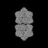 42232MC M: map data used to model this data C: citing same article ( |
|---|---|
| Similar structure data |
-Links
 PDBj
PDBj
- Assembly
| Deposited unit | 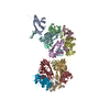
|
|---|---|
| 1 |
|
-Components
| #1: Protein | Mass: 26880.529 Da / Num. of mol.: 11 Source method: isolated from a genetically manipulated source Source: (gene. exp.)  Maize streak virus genotype A (isolate Nigeria) Maize streak virus genotype A (isolate Nigeria)Strain: isolate Nigeria / Gene: V1 / Production host:  Agrobacterium tumefaciens (bacteria) / References: UniProt: P06448 Agrobacterium tumefaciens (bacteria) / References: UniProt: P06448#2: DNA chain | Mass: 2669.778 Da / Num. of mol.: 11 Source method: isolated from a genetically manipulated source Source: (gene. exp.) Maize streak virus genotype A (isolate Nigeria)Strain: isolate Nigeria / Production host: Agrobacterium tumefaciens (bacteria) |
|---|
-Experimental details
-Experiment
| Experiment | Method: ELECTRON MICROSCOPY |
|---|---|
| EM experiment | Aggregation state: PARTICLE / 3D reconstruction method: single particle reconstruction |
- Sample preparation
| Component | Name: Maize streak virus - [Nigeria] / Type: VIRUS Details: Gemini capsid is a Pseudo T=1 icosahedron. The structure contains both protein and ssDNA. Entity ID: all / Source: RECOMBINANT |
|---|---|
| Source (natural) | Organism: Maize streak virus genotype A (isolate Nigeria) / Strain: isolate Nigeria |
| Source (recombinant) | Organism: Agrobacterium tumefaciens (bacteria) |
| Details of virus | Empty: NO / Enveloped: NO / Isolate: STRAIN / Type: VIRION |
| Natural host | Organism: Zea mays |
| Virus shell | Triangulation number (T number): 1 |
| Buffer solution | pH: 4 |
| Buffer component | Conc.: 0.1 M / Name: Sodium acetate / Formula: NaAc |
| Specimen | Conc.: 1 mg/ml / Embedding applied: NO / Shadowing applied: NO / Staining applied: NO / Vitrification applied: YES |
| Specimen support | Grid type: C-flat |
| Vitrification | Instrument: FEI VITROBOT MARK IV / Cryogen name: ETHANE |
- Electron microscopy imaging
| Experimental equipment |  Model: Titan Krios / Image courtesy: FEI Company |
|---|---|
| Microscopy | Model: FEI TITAN KRIOS |
| Electron gun | Electron source:  FIELD EMISSION GUN / Accelerating voltage: 300 kV / Illumination mode: SPOT SCAN FIELD EMISSION GUN / Accelerating voltage: 300 kV / Illumination mode: SPOT SCAN |
| Electron lens | Mode: BRIGHT FIELD / Nominal defocus max: 3627 nm / Nominal defocus min: 947 nm / Cs: 2.7 mm |
| Image recording | Electron dose: 60 e/Å2 / Film or detector model: DIRECT ELECTRON DE-64 (8k x 8k) / Num. of real images: 1408 |
- Processing
| EM software |
| ||||||||||||||||||||||||
|---|---|---|---|---|---|---|---|---|---|---|---|---|---|---|---|---|---|---|---|---|---|---|---|---|---|
| CTF correction | Type: PHASE FLIPPING AND AMPLITUDE CORRECTION | ||||||||||||||||||||||||
| Symmetry | Point symmetry: D5 (2x5 fold dihedral) | ||||||||||||||||||||||||
| 3D reconstruction | Resolution: 3.17 Å / Resolution method: FSC 0.143 CUT-OFF / Num. of particles: 1408 / Symmetry type: POINT | ||||||||||||||||||||||||
| Atomic model building | Protocol: AB INITIO MODEL | ||||||||||||||||||||||||
| Atomic model building | Type: in silico model | ||||||||||||||||||||||||
| Refine LS restraints |
|
