National Institutes of Health/National Institute of Diabetes and Digestive and Kidney Disease (NIH/NIDDK)
RO1 DK100605
United States
Citation
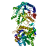
Journal: Sci Rep / Year: 2025 Title: Conformational landscape of soluble α-klotho revealed by cryogenic electron microscopy. Authors: Nicholas J Schnicker / Zhen Xu / Mohammad Amir / Lokesh Gakhar / Chou-Long Huang / Abstract: α-Klotho (KLA) is a type-1 membranous protein that can associate with fibroblast growth factor receptor (FGFR) to form co-receptor for FGF23. The ectodomain of unassociated KLA is shed as soluble ...α-Klotho (KLA) is a type-1 membranous protein that can associate with fibroblast growth factor receptor (FGFR) to form co-receptor for FGF23. The ectodomain of unassociated KLA is shed as soluble KLA (sKLA) to exert FGFR/FGF23-independent pleiotropic functions. The previously determined X-ray crystal structure of the extracellular region of sKLA in complex with FGF23 and FGFR1c suggests that sKLA functions solely as an on-demand coreceptor for FGF23. To understand the FGFR/FGF23-independent pleiotropic functions of sKLA, we investigated biophysical properties and structure of apo-sKLA. Single particle cryogenic electron microscopy (cryo-EM) revealed a 3.3 Å resolution structure of apo-sKLA that overlays well with its counterpart in the ternary complex with several distinct features. Compared to the ternary complex, the KL2 domain of apo-sKLA is more flexible. Three-dimensional variability analysis revealed that apo-sKLA adopts conformations with different KL1-KL2 interdomain bending and rotational angles. Mass photometry revealed that sKLA can form a stable structure with FGFR and/or FGF23 as well as sKLA dimer in solution. Cryo-EM supported the dimeric structure of sKLA. Recent studies revealed that FGF23 contains two KLA-binding sites. Our computational studies revealed that each site binds separate KLA in the dimer. The potential multiple forms and shapes of sKLA support its role as FGFR-independent hormone with pleiotropic functions. The ability of FGF23 to engage two KLA's simultaneously raises a potential new mechanism of action for FGF23-mediated signaling by the membranous klotho.
History
Deposition
Oct 3, 2023
Deposition site: RCSB / Processing site: RCSB
Revision 1.0
Oct 16, 2024
Provider: repository / Type: Initial release
Revision 1.0
Oct 16, 2024
Data content type: EM metadata / Data content type: EM metadata / Provider: repository / Type: Initial release
Revision 1.0
Oct 16, 2024
Data content type: FSC / Data content type: FSC / Provider: repository / Type: Initial release
Revision 1.0
Oct 16, 2024
Data content type: Half map / Part number: 1 / Data content type: Half map / Provider: repository / Type: Initial release
Revision 1.0
Oct 16, 2024
Data content type: Half map / Part number: 2 / Data content type: Half map / Provider: repository / Type: Initial release
Revision 1.0
Oct 16, 2024
Data content type: Image / Data content type: Image / Provider: repository / Type: Initial release
Revision 1.0
Oct 16, 2024
Data content type: Primary map / Data content type: Primary map / Provider: repository / Type: Initial release
Data content type: EM metadata / Data content type: EM metadata / EM metadata / Group: Database references / Experimental summary / Data content type: EM metadata / EM metadata / EM metadata / Category: citation / citation_author / em_admin Data content type: EM metadata / EM metadata ...EM metadata / EM metadata / EM metadata / EM metadata / EM metadata / EM metadata / EM metadata / EM metadata / EM metadata / EM metadata / EM metadata / EM metadata / EM metadata / EM metadata Item: _citation.country / _citation.journal_abbrev ..._citation.country / _citation.journal_abbrev / _citation.journal_id_CSD / _citation.journal_id_ISSN / _citation.journal_volume / _citation.page_first / _citation.page_last / _citation.pdbx_database_id_DOI / _citation.pdbx_database_id_PubMed / _citation.title / _citation.year / _citation_author.identifier_ORCID / _citation_author.name / _em_admin.last_update
Average exposure time: 2 sec. / Electron dose: 60 e/Å2 / Film or detector model: GATAN K3 (6k x 4k) / Num. of grids imaged: 1 / Num. of real images: 5760
-
Processing
EM software
ID
Name
Version
Category
4
cryoSPARC
3.3.1
CTFcorrection
7
UCSF Chimera
modelfitting
9
PHENIX
modelrefinement
10
cryoSPARC
3.3.1
initialEulerassignment
11
cryoSPARC
3.3.1
finalEulerassignment
CTF correction
Type: PHASE FLIPPING AND AMPLITUDE CORRECTION
Particle selection
Num. of particles selected: 7891498
3D reconstruction
Resolution: 6.5 Å / Resolution method: FSC 0.143 CUT-OFF / Num. of particles: 30219 / Symmetry type: POINT
In the structure databanks used in Yorodumi, some data are registered as the other names, "COVID-19 virus" and "2019-nCoV". Here are the details of the virus and the list of structure data.
Jan 31, 2019. EMDB accession codes are about to change! (news from PDBe EMDB page)
EMDB accession codes are about to change! (news from PDBe EMDB page)
The allocation of 4 digits for EMDB accession codes will soon come to an end. Whilst these codes will remain in use, new EMDB accession codes will include an additional digit and will expand incrementally as the available range of codes is exhausted. The current 4-digit format prefixed with “EMD-” (i.e. EMD-XXXX) will advance to a 5-digit format (i.e. EMD-XXXXX), and so on. It is currently estimated that the 4-digit codes will be depleted around Spring 2019, at which point the 5-digit format will come into force.
The EM Navigator/Yorodumi systems omit the EMD- prefix.
Related info.:Q: What is EMD? / ID/Accession-code notation in Yorodumi/EM Navigator
Yorodumi is a browser for structure data from EMDB, PDB, SASBDB, etc.
This page is also the successor to EM Navigator detail page, and also detail information page/front-end page for Omokage search.
The word "yorodu" (or yorozu) is an old Japanese word meaning "ten thousand". "mi" (miru) is to see.
Related info.:EMDB / PDB / SASBDB / Comparison of 3 databanks / Yorodumi Search / Aug 31, 2016. New EM Navigator & Yorodumi / Yorodumi Papers / Jmol/JSmol / Function and homology information / Changes in new EM Navigator and Yorodumi
 Movie
Movie Controller


 Open data
 Basic information
 Components
 Keywords
 Function and homology information
 Homo sapiens (human)
 Authors
 United States, 1items
  Citation
 Structure visualization
 Downloads & links
 Other downloads

 PDBj
PDBj

 Assembly

 Sample preparation
 Electron microscopy imaging
 FIELD EMISSION GUN / Accelerating voltage: 300 kV / Illumination mode: FLOOD BEAM
 Processing
