 ムービー
ムービー コントローラー


+ データを開く

- 基本情報
基本情報
| 登録情報 | データベース: PDB / ID: 8udi | ||||||
|---|---|---|---|---|---|---|---|
| タイトル | Crystal structure of Helicobacter pylori glutamate racemase bound to D-glutamate and a crystallographic artifact | ||||||
 要素 要素 | Glutamate racemase | ||||||
 キーワード キーワード | ISOMERASE / Enzyme / Epimerase / Gyrase / Artifiact | ||||||
| 機能・相同性 |  機能・相同性情報 機能・相同性情報glutamate racemase / glutamate racemase activity / peptidoglycan biosynthetic process / cell wall organization / regulation of cell shape 類似検索 - 分子機能 | ||||||
| 生物種 |   Helicobacter pylori (ピロリ菌) Helicobacter pylori (ピロリ菌) | ||||||
| 手法 |  X線回折 / シンクロトロン / 分子置換 / 解像度: 2.1 Å X線回折 / シンクロトロン / 分子置換 / 解像度: 2.1 Å | ||||||
 データ登録者 データ登録者 | Propp, J. / Spies, M.A. | ||||||
| 資金援助 |  米国, 1件 米国, 1件
| ||||||
 引用 引用 | ジャーナル: To Be Published タイトル: Structure of H. Pylori Glutamate Racemace with a crystallographic artifact 著者: Propp, J. / Spies, M.A. | ||||||
| 履歴 |
|
- 構造の表示
構造の表示
| 構造ビューア | 分子: MolmilJmol/JSmol |
|---|
- ダウンロードとリンク
-ダウンロード
| PDBx/mmCIF形式 | 8udi.cif.gz | 115.4 KB | 表示 | PDBx/mmCIF形式 |
|---|---|---|---|---|
| PDB形式 | pdb8udi.ent.gz | 86.5 KB | 表示 | PDB形式 |
| PDBx/mmJSON形式 | 8udi.json.gz | ツリー表示 | PDBx/mmJSON形式 | |
| その他 |  その他のダウンロード その他のダウンロード |
-検証レポート
| 文書・要旨 | 8udi_validation.pdf.gz | 766 KB | 表示 | wwPDB検証レポート |
|---|---|---|---|---|
| 文書・詳細版 | 8udi_full_validation.pdf.gz | 771 KB | 表示 | |
| XML形式データ | 8udi_validation.xml.gz | 24.6 KB | 表示 | |
| CIF形式データ | 8udi_validation.cif.gz | 32.6 KB | 表示 | |
| アーカイブディレクトリ | https://data.pdbj.org/pub/pdb/validation_reports/ud/8udiftp://data.pdbj.org/pub/pdb/validation_reports/ud/8udi | HTTPS FTP |
-関連構造データ
| 類似構造データ |
|---|
-リンク
 PDBj
PDBj- 集合体
集合体
| 登録構造単位 | 
| ||||||||||
|---|---|---|---|---|---|---|---|---|---|---|---|
| 1 |
| ||||||||||
| 単位格子 |
|
-要素
| #1: タンパク質 | 分子量: 28419.037 Da / 分子数: 2 / 由来タイプ: 組換発現 / 由来: (組換発現) Helicobacter pylori (ピロリ菌) / 遺伝子: murI / 発現宿主: #2: 化合物 | 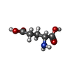  タイプ: D-peptide linking / 分子量: 147.129 Da / 分子数: 2 / 由来タイプ: 天然 / 式: C5H9NO4 タイプ: D-peptide linking / 分子量: 147.129 Da / 分子数: 2 / 由来タイプ: 天然 / 式: C5H9NO4#3: 化合物 | ChemComp-WAW / | 分子量: 402.424 Da / 分子数: 1 / 由来タイプ: 合成 / 式: C22H19FN6O / タイプ: SUBJECT OF INVESTIGATION #4: 化合物 |   分子量: 35.453 Da / 分子数: 2 / 由来タイプ: 合成 / 式: Cl 分子量: 35.453 Da / 分子数: 2 / 由来タイプ: 合成 / 式: Cl#5: 水 | ChemComp-HOH / |  分子量: 18.015 Da / 分子数: 168 / 由来タイプ: 天然 / 式: H2O 分子量: 18.015 Da / 分子数: 168 / 由来タイプ: 天然 / 式: H2O研究の焦点であるリガンドがあるか | Y | Has protein modification | N | |
|---|
-実験情報
-実験
| 実験 | 手法: X線回折 / 使用した結晶の数: 1 |
|---|
- 試料調製
試料調製
| 結晶 | マシュー密度: 2.58 Å3/Da / 溶媒含有率: 52.31 % |
|---|---|
| 結晶化 | 温度: 293 K / 手法: 蒸気拡散法, ハンギングドロップ法 詳細: 4.5g/l HpGR 200mM Ammonium Acetate, 5mM D/L-Glutamate 20% DMSO 2.5mM ligand mixed 2:1 with INDEX G1 8% DMSO 1mM M3 |
-データ収集
| 回折 | 平均測定温度: 100 K / Serial crystal experiment: N |
|---|---|
| 放射光源 | 由来: シンクロトロン / サイト: APS / ビームライン: 24-ID-E / 波長: 0.9272 Å |
| 検出器 | タイプ: DECTRIS EIGER X 16M / 検出器: PIXEL / 日付: 2023年2月18日 |
| 放射 | プロトコル: SINGLE WAVELENGTH / 単色(M)・ラウエ(L): M / 散乱光タイプ: x-ray |
| 放射波長 | 波長: 0.9272 Å / 相対比: 1 |
| 反射 | 解像度: 2.1→81.97 Å / Num. obs: 34681 / % possible obs: 99.7 % / 冗長度: 5.2 % / Biso Wilson estimate: 42.51 Å2 / CC1/2: 0.999 / Rmerge(I) obs: 0.042 / Rpim(I) all: 0.02 / Rrim(I) all: 0.047 / Χ2: 0.95 / Net I/σ(I): 22.6 / Num. measured all: 180639 |
| 反射 シェル | 解像度: 2.1→2.16 Å / % possible obs: 99.9 % / 冗長度: 5.5 % / Rmerge(I) obs: 0.523 / Num. measured all: 15389 / Num. unique obs: 2818 / CC1/2: 0.906 / Rpim(I) all: 0.24 / Rrim(I) all: 0.577 / Χ2: 0.76 / Net I/σ(I) obs: 3.9 |
- 解析
解析
| ソフトウェア |
| |||||||||||||||||||||||||||||||||||||||||||||||||||||||||||||||||||||||||||||||||||||||||||
|---|---|---|---|---|---|---|---|---|---|---|---|---|---|---|---|---|---|---|---|---|---|---|---|---|---|---|---|---|---|---|---|---|---|---|---|---|---|---|---|---|---|---|---|---|---|---|---|---|---|---|---|---|---|---|---|---|---|---|---|---|---|---|---|---|---|---|---|---|---|---|---|---|---|---|---|---|---|---|---|---|---|---|---|---|---|---|---|---|---|---|---|---|
| 精密化 | 構造決定の手法: 分子置換 / 解像度: 2.1→66.54 Å / SU ML: 0.235 / 交差検証法: FREE R-VALUE / σ(F): 1.35 / 位相誤差: 25.0366 立体化学のターゲット値: GeoStd + Monomer Library + CDL v1.2
| |||||||||||||||||||||||||||||||||||||||||||||||||||||||||||||||||||||||||||||||||||||||||||
| 溶媒の処理 | 減衰半径: 0.9 Å / VDWプローブ半径: 1.1 Å / 溶媒モデル: FLAT BULK SOLVENT MODEL | |||||||||||||||||||||||||||||||||||||||||||||||||||||||||||||||||||||||||||||||||||||||||||
| 原子変位パラメータ | Biso mean: 47.34 Å2 | |||||||||||||||||||||||||||||||||||||||||||||||||||||||||||||||||||||||||||||||||||||||||||
| 精密化ステップ | サイクル: LAST / 解像度: 2.1→66.54 Å
| |||||||||||||||||||||||||||||||||||||||||||||||||||||||||||||||||||||||||||||||||||||||||||
| 拘束条件 |
| |||||||||||||||||||||||||||||||||||||||||||||||||||||||||||||||||||||||||||||||||||||||||||
| LS精密化 シェル |
|
