 Movie
Movie Controller


+ Open data

- Basic information
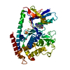
| Entry | Database: PDB / ID: 8uct | |||||||||
|---|---|---|---|---|---|---|---|---|---|---|
| Title | Crystal structure of TcPINK1 in complex with PRT | |||||||||
 Components Components | Serine/threonine-protein kinase Pink1, mitochondrial | |||||||||
 Keywords Keywords | CELL CYCLE / Mitophagy Autophagy Kinase | |||||||||
| Function / homology |  Function and homology information Function and homology informationpositive regulation of free ubiquitin chain polymerization / autophagy of mitochondrion / positive regulation of mitochondrial fission / positive regulation of protein ubiquitination / protein autophosphorylation / regulation of apoptotic process / mitochondrial outer membrane / non-specific serine/threonine protein kinase / mitochondrial inner membrane / protein serine/threonine kinase activity ...positive regulation of free ubiquitin chain polymerization / autophagy of mitochondrion / positive regulation of mitochondrial fission / positive regulation of protein ubiquitination / protein autophosphorylation / regulation of apoptotic process / mitochondrial outer membrane / non-specific serine/threonine protein kinase / mitochondrial inner membrane / protein serine/threonine kinase activity / ubiquitin protein ligase binding / mitochondrion / ATP binding / metal ion binding / cytosol Similarity search - Function | |||||||||
| Biological species |  | |||||||||
| Method |  X-RAY DIFFRACTION / SYNCHROTRON / MOLECULAR REPLACEMENT / Resolution: 2.93 Å X-RAY DIFFRACTION / SYNCHROTRON / MOLECULAR REPLACEMENT / Resolution: 2.93 Å | |||||||||
 Authors Authors | Veyron, S. / Rasool, S. / Trempe, J.F. | |||||||||
| Funding support |  Canada, Canada,  United States, 2items United States, 2items
| |||||||||
 Citation Citation | Journal: Sci Rep / Year: 2024 Title: Identification and structural characterization of small molecule inhibitors of PINK1. Authors: Rasool, S. / Shomali, T. / Truong, L. / Croteau, N. / Veyron, S. / Bustillos, B.A. / Springer, W. / Fiesel, F.C. / Trempe, J.F. | |||||||||
| History |
|
- Structure visualization
| Structure viewer | Molecule: MolmilJmol/JSmol |
|---|
- Downloads & links
-Download
| PDBx/mmCIF format | 8uct.cif.gz | 256.6 KB | Display | PDBx/mmCIF format |
|---|---|---|---|---|
| PDB format | pdb8uct.ent.gz | 185.3 KB | Display | PDB format |
| PDBx/mmJSON format | 8uct.json.gz | Tree view | PDBx/mmJSON format | |
| Others |  Other downloads Other downloads |
-Validation report
| Arichive directory | https://data.pdbj.org/pub/pdb/validation_reports/uc/8uctftp://data.pdbj.org/pub/pdb/validation_reports/uc/8uct | HTTPS FTP |
|---|
-Related structure data
-Links
 PDBj
PDBj


- Assembly
| Deposited unit | 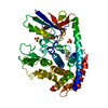
| ||||||||||||
|---|---|---|---|---|---|---|---|---|---|---|---|---|---|
| 1 | 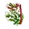
| ||||||||||||
| Unit cell |
|
-Components
| #1: Protein | Mass: 51032.293 Da / Num. of mol.: 1 / Mutation: W131A, W142A, Y225A, Y378A, F401A, F437A Source method: isolated from a genetically manipulated source Source: (gene. exp.) Production host:  References: UniProt: D6WMX4 | ||||||||
|---|---|---|---|---|---|---|---|---|---|
| #2: Chemical | ChemComp-DTT /   Mass: 154.251 Da / Num. of mol.: 1 / Source method: obtained synthetically / Formula: C4H10O2S2 Mass: 154.251 Da / Num. of mol.: 1 / Source method: obtained synthetically / Formula: C4H10O2S2 | ||||||||
| #3: Chemical |   Mass: 96.063 Da / Num. of mol.: 2 / Source method: isolated from a natural source / Formula: SO4 Mass: 96.063 Da / Num. of mol.: 2 / Source method: isolated from a natural source / Formula: SO4#4: Chemical | ChemComp-3YT / | 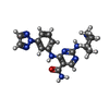  Mass: 393.446 Da / Num. of mol.: 1 / Source method: obtained synthetically / Formula: C19H23N9O / Feature type: SUBJECT OF INVESTIGATION Mass: 393.446 Da / Num. of mol.: 1 / Source method: obtained synthetically / Formula: C19H23N9O / Feature type: SUBJECT OF INVESTIGATION#5: Water | ChemComp-HOH / |  Mass: 18.015 Da / Num. of mol.: 23 / Source method: isolated from a natural source / Formula: H2O Mass: 18.015 Da / Num. of mol.: 23 / Source method: isolated from a natural source / Formula: H2OHas ligand of interest | Y | Has protein modification | N | |
-Experimental details
-Experiment
| Experiment | Method: X-RAY DIFFRACTION / Number of used crystals: 1 |
|---|
- Sample preparation
| Crystal | Density Matthews: 2.18 Å3/Da / Density % sol: 43.71 % |
|---|---|
| Crystal grow | Temperature: 295 K / Method: vapor diffusion, hanging drop / pH: 7 / Details: 100mM HEPES, PEG 4000, 150mM ammonium sulfate |
-Data collection
| Diffraction | Mean temperature: 100 K / Serial crystal experiment: N |
|---|---|
| Diffraction source | Source: SYNCHROTRON / Site: APS / Beamline: 24-ID-C / Wavelength: 0.987 Å |
| Detector | Type: DECTRIS EIGER2 X 16M / Detector: PIXEL / Date: Nov 27, 2021 |
| Radiation | Protocol: SINGLE WAVELENGTH / Monochromatic (M) / Laue (L): M / Scattering type: x-ray |
| Radiation wavelength | Wavelength: 0.987 Å / Relative weight: 1 |
| Reflection | Resolution: 2.93→90.93 Å / Num. obs: 11014 / % possible obs: 100 % / Redundancy: 17.8 % / Biso Wilson estimate: 75.42 Å2 / CC1/2: 0.995 / Rpim(I) all: 0.145 / Net I/σ(I): 6.1 |
| Reflection shell | Resolution: 2.93→2.98 Å / Redundancy: 16.9 % / Mean I/σ(I) obs: 0.5 / Num. unique obs: 521 / CC1/2: 0.551 / % possible all: 100 |
- Processing
| Software |
| |||||||||||||||||||||||||||||||||||
|---|---|---|---|---|---|---|---|---|---|---|---|---|---|---|---|---|---|---|---|---|---|---|---|---|---|---|---|---|---|---|---|---|---|---|---|---|
| Refinement | Method to determine structure: MOLECULAR REPLACEMENT / Resolution: 2.93→90.93 Å / SU ML: 0.4527 / Cross valid method: FREE R-VALUE / σ(F): 1.34 / Phase error: 30.0372 Stereochemistry target values: GeoStd + Monomer Library + CDL v1.2
| |||||||||||||||||||||||||||||||||||
| Solvent computation | Shrinkage radii: 0.9 Å / VDW probe radii: 1.1 Å / Solvent model: FLAT BULK SOLVENT MODEL | |||||||||||||||||||||||||||||||||||
| Displacement parameters | Biso mean: 76.82 Å2 | |||||||||||||||||||||||||||||||||||
| Refinement step | Cycle: LAST / Resolution: 2.93→90.93 Å
| |||||||||||||||||||||||||||||||||||
| Refine LS restraints |
| |||||||||||||||||||||||||||||||||||
| LS refinement shell |
|
