 Movie
Movie Controller

[English] 日本語
 Yorodumi
- PDB-8uc4: Apo X-ray crystal structure of Cyclophilin D with a surface entro... -
+ Open data

- Basic information
| Entry | Database: PDB / ID: 8uc4 | ||||||||||||
|---|---|---|---|---|---|---|---|---|---|---|---|---|---|
| Title | Apo X-ray crystal structure of Cyclophilin D with a surface entropy reduction mutation (K175I) | ||||||||||||
 Components Components | Peptidyl-prolyl cis-trans isomerase F, mitochondrial | ||||||||||||
 Keywords Keywords | ISOMERASE / Cyclophilin D / mitochondria | ||||||||||||
| Function / homology |  Function and homology information Function and homology information: / mitochondrial outer membrane permeabilization involved in programmed cell death / regulation of mitochondrial membrane permeability involved in programmed necrotic cell death / skeletal muscle fiber differentiation / mitochondrial permeability transition pore complex / cellular response to arsenic-containing substance / mitochondrial depolarization / negative regulation of oxidative phosphorylation / regulation of mitochondrial membrane permeability / cyclosporin A binding ...: / mitochondrial outer membrane permeabilization involved in programmed cell death / regulation of mitochondrial membrane permeability involved in programmed necrotic cell death / skeletal muscle fiber differentiation / mitochondrial permeability transition pore complex / cellular response to arsenic-containing substance / mitochondrial depolarization / negative regulation of oxidative phosphorylation / regulation of mitochondrial membrane permeability / cyclosporin A binding / negative regulation of release of cytochrome c from mitochondria / necroptotic process / negative regulation of intrinsic apoptotic signaling pathway / apoptotic mitochondrial changes / cellular response to calcium ion / response to ischemia / peptidylprolyl isomerase / peptidyl-prolyl cis-trans isomerase activity / cellular response to hydrogen peroxide / protein folding / mitochondrial matrix / negative regulation of apoptotic process / mitochondrion / membrane / cytoplasm Similarity search - Function | ||||||||||||
| Biological species |  Homo sapiens (human) Homo sapiens (human) | ||||||||||||
| Method |  X-RAY DIFFRACTION / SYNCHROTRON / MOLECULAR REPLACEMENT / Resolution: 1.87 Å X-RAY DIFFRACTION / SYNCHROTRON / MOLECULAR REPLACEMENT / Resolution: 1.87 Å | ||||||||||||
 Authors Authors | Kreitler, D.F. / Rangwala, A.M. / Seeliger, M.A. | ||||||||||||
| Funding support |  United States, 3items United States, 3items
| ||||||||||||
 Citation Citation | Journal: To Be Published Title: Apo X-ray crystal structure of Cyclophilin D with a surface entropy reduction mutation (K175I) Authors: Kreitler, D.F. / Seeliger, M.A. / Rangwala, A.M. | ||||||||||||
| History |
|
- Structure visualization
| Structure viewer | Molecule: MolmilJmol/JSmol |
|---|
- Downloads & links
-Download
| PDBx/mmCIF format | 8uc4.cif.gz | 57.8 KB | Display | PDBx/mmCIF format |
|---|---|---|---|---|
| PDB format | pdb8uc4.ent.gz | 33.2 KB | Display | PDB format |
| PDBx/mmJSON format | 8uc4.json.gz | Tree view | PDBx/mmJSON format | |
| Others |  Other downloads Other downloads |
-Validation report
| Arichive directory | https://data.pdbj.org/pub/pdb/validation_reports/uc/8uc4ftp://data.pdbj.org/pub/pdb/validation_reports/uc/8uc4 | HTTPS FTP |
|---|
-Related structure data

-Links
 PDBj
PDBj

- Assembly
| Deposited unit | 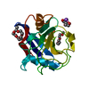
| ||||||||||||
|---|---|---|---|---|---|---|---|---|---|---|---|---|---|
| 1 |
| ||||||||||||
| Unit cell |
| ||||||||||||
| Components on special symmetry positions |
|
-Components
| #1: Protein | Mass: 17867.334 Da / Num. of mol.: 1 / Mutation: K175I Source method: isolated from a genetically manipulated source Source: (gene. exp.) Homo sapiens (human) / Gene: PPIF, CYP3 / Production host:  | ||||||||
|---|---|---|---|---|---|---|---|---|---|
| #2: Chemical |   Mass: 1529.829 Da / Num. of mol.: 2 / Source method: obtained synthetically / Formula: C69H140O35 / Comment: precipitant*YM Mass: 1529.829 Da / Num. of mol.: 2 / Source method: obtained synthetically / Formula: C69H140O35 / Comment: precipitant*YM#3: Chemical |   Mass: 106.120 Da / Num. of mol.: 2 / Source method: obtained synthetically / Formula: C4H10O3 Mass: 106.120 Da / Num. of mol.: 2 / Source method: obtained synthetically / Formula: C4H10O3#4: Chemical | ChemComp-NA / |   Mass: 22.990 Da / Num. of mol.: 1 / Source method: obtained synthetically / Formula: Na Mass: 22.990 Da / Num. of mol.: 1 / Source method: obtained synthetically / Formula: Na#5: Water | ChemComp-HOH / |  Mass: 18.015 Da / Num. of mol.: 119 / Source method: isolated from a natural source / Formula: H2O Mass: 18.015 Da / Num. of mol.: 119 / Source method: isolated from a natural source / Formula: H2OHas ligand of interest | N | |
-Experimental details
-Experiment
| Experiment | Method: X-RAY DIFFRACTION / Number of used crystals: 1 |
|---|
- Sample preparation
| Crystal | Density Matthews: 2.49 Å3/Da / Density % sol: 50.64 % / Description: 3D |
|---|---|
| Crystal grow | Temperature: 293 K / Method: vapor diffusion, hanging drop / pH: 7 Details: Well solution: 40% w/v PEG3350, 30 mM NaCl, 50 mM potassium phosphate monobasic pH 7.0 Protein Solution: 15 mg/mL protein, 20 mM Tris pH 8.0, 50 mM NaCl, 1 mM DTT, and 5% glycerol Drop: 1 uL ...Details: Well solution: 40% w/v PEG3350, 30 mM NaCl, 50 mM potassium phosphate monobasic pH 7.0 Protein Solution: 15 mg/mL protein, 20 mM Tris pH 8.0, 50 mM NaCl, 1 mM DTT, and 5% glycerol Drop: 1 uL protein solution, 1 uL well solution |
-Data collection
| Diffraction | Mean temperature: 100 K / Serial crystal experiment: N |
|---|---|
| Diffraction source | Source: SYNCHROTRON / Site: NSLS-II / Beamline: 17-ID-1 / Wavelength: 0.9201 Å |
| Detector | Type: DECTRIS EIGER X 9M / Detector: PIXEL / Date: Feb 18, 2022 / Details: KB |
| Radiation | Monochromator: DCM Si(111) / Protocol: SINGLE WAVELENGTH / Monochromatic (M) / Laue (L): M / Scattering type: x-ray |
| Radiation wavelength | Wavelength: 0.9201 Å / Relative weight: 1 |
| Reflection | Resolution: 1.87→28.48 Å / Num. obs: 15443 / % possible obs: 99.2 % / Redundancy: 7.2 % / Biso Wilson estimate: 23.25 Å2 / CC1/2: 0.996 / Rrim(I) all: 0.137 / Net I/σ(I): 9.8 |
| Reflection shell | Resolution: 1.87→1.92 Å / Redundancy: 6.4 % / Num. unique obs: 1014 / CC1/2: 0.821 / % possible all: 90.2 |
- Processing
| Software |
| ||||||||||||||||||||||||||||||||||||||||||||||||||||||||||||||||||||||||||||||||||||
|---|---|---|---|---|---|---|---|---|---|---|---|---|---|---|---|---|---|---|---|---|---|---|---|---|---|---|---|---|---|---|---|---|---|---|---|---|---|---|---|---|---|---|---|---|---|---|---|---|---|---|---|---|---|---|---|---|---|---|---|---|---|---|---|---|---|---|---|---|---|---|---|---|---|---|---|---|---|---|---|---|---|---|---|---|---|
| Refinement | Method to determine structure: MOLECULAR REPLACEMENT / Resolution: 1.87→28.48 Å / SU ML: 0.1751 / Cross valid method: FREE R-VALUE / σ(F): 1.35 / Phase error: 19.9029 Stereochemistry target values: GeoStd + Monomer Library + CDL v1.2
| ||||||||||||||||||||||||||||||||||||||||||||||||||||||||||||||||||||||||||||||||||||
| Solvent computation | Shrinkage radii: 0.9 Å / VDW probe radii: 1.11 Å / Solvent model: FLAT BULK SOLVENT MODEL | ||||||||||||||||||||||||||||||||||||||||||||||||||||||||||||||||||||||||||||||||||||
| Displacement parameters | Biso mean: 25.14 Å2 | ||||||||||||||||||||||||||||||||||||||||||||||||||||||||||||||||||||||||||||||||||||
| Refinement step | Cycle: LAST / Resolution: 1.87→28.48 Å
| ||||||||||||||||||||||||||||||||||||||||||||||||||||||||||||||||||||||||||||||||||||
| Refine LS restraints |
| ||||||||||||||||||||||||||||||||||||||||||||||||||||||||||||||||||||||||||||||||||||
| LS refinement shell |
|
