 Movie
Movie Controller

[English] 日本語
 Yorodumi
- PDB-8uc3: Cryo-EM structure of the AlbAB cyclodipeptide oxidase enzyme filament -
+ Open data

- Basic information
| Entry | Database: PDB / ID: 8uc3 | ||||||
|---|---|---|---|---|---|---|---|
| Title | Cryo-EM structure of the AlbAB cyclodipeptide oxidase enzyme filament | ||||||
 Components Components |
| ||||||
 Keywords Keywords | OXIDOREDUCTASE / cyclodipeptide oxidase / cyclic dipeptide oxidase / nitroreductase-like / enzyme filament / flavoenzyme | ||||||
| Function / homology |  Function and homology information Function and homology informationalbonoursin synthase / oxidoreductase activity, acting on the CH-CH group of donors, oxygen as acceptor / cytoplasm Similarity search - Function | ||||||
| Biological species |  Streptomyces noursei ATCC 11455 (bacteria) Streptomyces noursei ATCC 11455 (bacteria) | ||||||
| Method | ELECTRON MICROSCOPY / helical reconstruction / cryo EM / Resolution: 2.78 Å | ||||||
 Authors Authors | Andreas, M.P. / Giessen, T.W. | ||||||
| Funding support |  United States, 1items United States, 1items
| ||||||
 Citation Citation | Journal: Nat Commun / Year: 2024 Title: Cyclodipeptide oxidase is an enzyme filament. Authors: Michael P Andreas / Tobias W Giessen / Abstract: Modified cyclic dipeptides represent a widespread class of secondary metabolites with diverse pharmacological activities, including antibacterial, antifungal, and antitumor. Here, we report the ...Modified cyclic dipeptides represent a widespread class of secondary metabolites with diverse pharmacological activities, including antibacterial, antifungal, and antitumor. Here, we report the structural characterization of the Streptomyces noursei enzyme AlbAB, a cyclodipeptide oxidase (CDO) carrying out α,β-dehydrogenations during the biosynthesis of the antibiotic albonoursin. We show that AlbAB is a megadalton heterooligomeric enzyme filament containing covalently bound flavin mononucleotide cofactors. We highlight that AlbAB filaments consist of alternating dimers of AlbA and AlbB and that enzyme activity is crucially dependent on filament formation. We show that AlbA-AlbB interactions are highly conserved suggesting that other CDO-like enzymes are likely enzyme filaments. As CDOs have been employed in the structural diversification of cyclic dipeptides, our results will be useful for future applications of CDOs in biocatalysis and chemoenzymatic synthesis. | ||||||
| History |
|
- Structure visualization
| Structure viewer | Molecule: MolmilJmol/JSmol |
|---|
- Downloads & links
-Download
| PDBx/mmCIF format | 8uc3.cif.gz | 117.8 KB | Display | PDBx/mmCIF format |
|---|---|---|---|---|
| PDB format | pdb8uc3.ent.gz | 90.5 KB | Display | PDB format |
| PDBx/mmJSON format | 8uc3.json.gz | Tree view | PDBx/mmJSON format | |
| Others |  Other downloads Other downloads |
-Validation report
| Summary document | 8uc3_validation.pdf.gz | 1.4 MB | Display | wwPDB validaton report |
|---|---|---|---|---|
| Full document | 8uc3_full_validation.pdf.gz | 1.5 MB | Display | |
| Data in XML | 8uc3_validation.xml.gz | 37 KB | Display | |
| Data in CIF | 8uc3_validation.cif.gz | 51.3 KB | Display | |
| Arichive directory | https://data.pdbj.org/pub/pdb/validation_reports/uc/8uc3ftp://data.pdbj.org/pub/pdb/validation_reports/uc/8uc3 | HTTPS FTP |
-Related structure data
| Related structure data |  42114MC M: map data used to model this data C: citing same article ( |
|---|---|
| Similar structure data |
-Links
 PDBj
PDBj- Assembly
| Deposited unit | 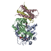
|
|---|---|
| 1 |
|
-Components
| #1: Protein | Mass: 21071.307 Da / Num. of mol.: 2 Source method: isolated from a genetically manipulated source Source: (gene. exp.) Streptomyces noursei ATCC 11455 (bacteria)Gene: albA / Production host: Streptomyces coelicolor A3(2) (bacteria) / Strain (production host): John Innes Centre M145 / References: UniProt: Q8GED9#2: Protein | Mass: 11583.143 Da / Num. of mol.: 2 Source method: isolated from a genetically manipulated source Source: (gene. exp.) Streptomyces noursei ATCC 11455 (bacteria)Gene: albB / Production host: Streptomyces coelicolor A3(2) (bacteria) / Strain (production host): John Innes Centre M145 / References: UniProt: Q8GED8#3: Chemical |   Mass: 456.344 Da / Num. of mol.: 2 / Source method: obtained synthetically / Formula: C17H21N4O9P / Feature type: SUBJECT OF INVESTIGATION Mass: 456.344 Da / Num. of mol.: 2 / Source method: obtained synthetically / Formula: C17H21N4O9P / Feature type: SUBJECT OF INVESTIGATIONHas ligand of interest | Y | Has protein modification | Y | |
|---|
-Experimental details
-Experiment
| Experiment | Method: ELECTRON MICROSCOPY |
|---|---|
| EM experiment | Aggregation state: FILAMENT / 3D reconstruction method: helical reconstruction |
- Sample preparation
| Component | Name: AlbAB cyclodipeptide oxidase enzyme filament / Type: COMPLEX / Entity ID: #1-#2 / Source: RECOMBINANT | |||||||||||||||
|---|---|---|---|---|---|---|---|---|---|---|---|---|---|---|---|---|
| Source (natural) | Organism: Streptomyces noursei ATCC 11455 (bacteria) | |||||||||||||||
| Source (recombinant) | Organism: Streptomyces coelicolor A3(2) (bacteria) / Strain: John Innes Centre M145 | |||||||||||||||
| Buffer solution | pH: 7.5 / Details: 20 mM NaCl, 150 mM Tris pH 7.5 | |||||||||||||||
| Buffer component |
| |||||||||||||||
| Specimen | Conc.: 0.75 mg/ml / Embedding applied: NO / Shadowing applied: NO / Staining applied: NO / Vitrification applied: YES | |||||||||||||||
| Specimen support | Details: Grid was glow discharged for 60 seconds at 5 mA under vacuum Grid material: COPPER / Grid mesh size: 200 divisions/in. / Grid type: Quantifoil R2/1 | |||||||||||||||
| Vitrification | Instrument: FEI VITROBOT MARK IV / Cryogen name: ETHANE / Humidity: 100 % / Chamber temperature: 295 K Details: Grid was plunge frozen into liquid ethane using the following parameters: blot force- 5, blot time 2 seconds |
- Electron microscopy imaging
| Experimental equipment |  Model: Titan Krios / Image courtesy: FEI Company |
|---|---|
| Microscopy | Model: FEI TITAN KRIOS |
| Electron gun | Electron source:  FIELD EMISSION GUN / Accelerating voltage: 300 kV / Illumination mode: FLOOD BEAM FIELD EMISSION GUN / Accelerating voltage: 300 kV / Illumination mode: FLOOD BEAM |
| Electron lens | Mode: BRIGHT FIELD / Nominal magnification: 105000 X / Nominal defocus max: 2500 nm / Nominal defocus min: 1000 nm / Cs: 2.7 mm / C2 aperture diameter: 100 µm |
| Specimen holder | Cryogen: NITROGEN / Specimen holder model: FEI TITAN KRIOS AUTOGRID HOLDER |
| Image recording | Average exposure time: 2.03063 sec. / Electron dose: 50.12 e/Å2 / Film or detector model: GATAN K3 BIOQUANTUM (6k x 4k) / Num. of grids imaged: 1 / Num. of real images: 3015 |
| Image scans | Width: 4092 / Height: 5760 |
- Processing
| EM software |
| ||||||||||||||||||||||||||||||||||||
|---|---|---|---|---|---|---|---|---|---|---|---|---|---|---|---|---|---|---|---|---|---|---|---|---|---|---|---|---|---|---|---|---|---|---|---|---|---|
| CTF correction | Type: PHASE FLIPPING AND AMPLITUDE CORRECTION | ||||||||||||||||||||||||||||||||||||
| Helical symmerty | Angular rotation/subunit: 119.969 ° / Axial rise/subunit: 46.063 Å / Axial symmetry: C1 | ||||||||||||||||||||||||||||||||||||
| Particle selection | Num. of particles selected: 1202923 | ||||||||||||||||||||||||||||||||||||
| 3D reconstruction | Resolution: 2.78 Å / Resolution method: FSC 0.143 CUT-OFF / Num. of particles: 795765 Details: Final reconstruction was generated by a masked local refinement of the helically refined map. The mask encompassed one dimer of AlbA and two surrounding dimers of AlbB. Symmetry type: HELICAL | ||||||||||||||||||||||||||||||||||||
| Atomic model building | B value: 107.4 / Protocol: FLEXIBLE FIT / Space: REAL / Target criteria: Cross-correlation coefficient Details: A model of AlbAB was created using AlphaFold2 using MMseqs2 via ColabFold. The model was then fit into the cryoEM density using ChimeraX. The model was then iteratively refined using Coot v ...Details: A model of AlbAB was created using AlphaFold2 using MMseqs2 via ColabFold. The model was then fit into the cryoEM density using ChimeraX. The model was then iteratively refined using Coot v 0.9.8.1 and real-space refinement in Phenix v 1.20.1-4487. | ||||||||||||||||||||||||||||||||||||
| Atomic model building | Source name: AlphaFold / Type: in silico model |
