 Movie
Movie Controller

[English] 日本語
 Yorodumi
- PDB-8ua5: Crystal Structure of infected cell protein 0 (ICP0) from herpes s... -
+ Open data

- Basic information
| Entry | Database: PDB / ID: 8ua5 | |||||||||
|---|---|---|---|---|---|---|---|---|---|---|
| Title | Crystal Structure of infected cell protein 0 (ICP0) from herpes simplex virus 1 (A636-Q776) | |||||||||
 Components Components | RL2 | |||||||||
 Keywords Keywords | PROTEIN BINDING / infected cell protein 0 (ICP0) / herpes simplex virus 1 / beta barrel / dimization | |||||||||
| Function / homology |  Function and homology information Function and homology informationligase activity / ubiquitin protein ligase activity / symbiont-mediated perturbation of host ubiquitin-like protein modification / protein ubiquitination / zinc ion binding Similarity search - Function | |||||||||
| Biological species |   Human alphaherpesvirus 1 (Herpes simplex virus type 1) Human alphaherpesvirus 1 (Herpes simplex virus type 1) | |||||||||
| Method |  X-RAY DIFFRACTION / SYNCHROTRON / MOLECULAR REPLACEMENT / Resolution: 2.45 Å X-RAY DIFFRACTION / SYNCHROTRON / MOLECULAR REPLACEMENT / Resolution: 2.45 Å | |||||||||
 Authors Authors | Lovell, S. / Kashipathy, M. / Battaile, K.P. / Cooper, A. / Davido, D. | |||||||||
| Funding support |  United States, 2items United States, 2items
| |||||||||
 Citation Citation | Journal: Proteins / Year: 2024 Title: HSV-1 ICP0 dimer domain adopts a novel beta-barrel fold. Authors: McCloskey, E. / Kashipathy, M. / Cooper, A. / Gao, P. / Johnson, D.K. / Battaile, K.P. / Lovell, S. / Davido, D.J. | |||||||||
| History |
|
- Structure visualization
| Structure viewer | Molecule: MolmilJmol/JSmol |
|---|
- Downloads & links
-Download
| PDBx/mmCIF format | 8ua5.cif.gz | 60 KB | Display | PDBx/mmCIF format |
|---|---|---|---|---|
| PDB format | pdb8ua5.ent.gz | 41.3 KB | Display | PDB format |
| PDBx/mmJSON format | 8ua5.json.gz | Tree view | PDBx/mmJSON format | |
| Others |  Other downloads Other downloads |
-Validation report
| Arichive directory | https://data.pdbj.org/pub/pdb/validation_reports/ua/8ua5ftp://data.pdbj.org/pub/pdb/validation_reports/ua/8ua5 | HTTPS FTP |
|---|
-Related structure data

-Links
 PDBj
PDBj

- Assembly
| Deposited unit | 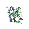
| ||||||||
|---|---|---|---|---|---|---|---|---|---|
| 1 | 
| ||||||||
| Unit cell |
|
-Components
| #1: Protein | Mass: 17842.926 Da / Num. of mol.: 2 / Fragment: A636-Q776 Source method: isolated from a genetically manipulated source Source: (gene. exp.) Human alphaherpesvirus 1 (Herpes simplex virus type 1)Gene: RL2, RL2_1, HHV1gp002, HHV1gp082 / Plasmid: pTBSG / Production host:  #2: Chemical | ChemComp-CL / |   Mass: 35.453 Da / Num. of mol.: 1 / Source method: obtained synthetically / Formula: Cl Mass: 35.453 Da / Num. of mol.: 1 / Source method: obtained synthetically / Formula: Cl#3: Chemical |   Mass: 126.904 Da / Num. of mol.: 3 / Source method: obtained synthetically / Formula: I Mass: 126.904 Da / Num. of mol.: 3 / Source method: obtained synthetically / Formula: I#4: Chemical | ChemComp-GOL / |   Mass: 92.094 Da / Num. of mol.: 1 / Source method: obtained synthetically / Formula: C3H8O3 Mass: 92.094 Da / Num. of mol.: 1 / Source method: obtained synthetically / Formula: C3H8O3#5: Water | ChemComp-HOH / |  Mass: 18.015 Da / Num. of mol.: 42 / Source method: isolated from a natural source / Formula: H2O Mass: 18.015 Da / Num. of mol.: 42 / Source method: isolated from a natural source / Formula: H2OHas ligand of interest | N | |
|---|
-Experimental details
-Experiment
| Experiment | Method: X-RAY DIFFRACTION / Number of used crystals: 1 |
|---|
- Sample preparation
| Crystal | Density Matthews: 2.54 Å3/Da / Density % sol: 51.51 % |
|---|---|
| Crystal grow | Temperature: 291 K / Method: vapor diffusion, sitting drop / pH: 7.5 Details: PACT G3: 20% (w/v) PEG 3350, 100 mM Bis-Tris Propane pH 7.5, 200 mM NaI, 80% crystallization solution and 20% (v/v) PEG 200. |
-Data collection
| Diffraction | Mean temperature: 100 K / Serial crystal experiment: N |
|---|---|
| Diffraction source | Source: SYNCHROTRON / Site: APS / Beamline: 17-ID / Wavelength: 1 Å |
| Detector | Type: DECTRIS PILATUS 6M / Detector: PIXEL / Date: Jun 15, 2019 |
| Radiation | Monochromator: Double Crystal Si 111 / Protocol: SINGLE WAVELENGTH / Monochromatic (M) / Laue (L): M / Scattering type: x-ray |
| Radiation wavelength | Wavelength: 1 Å / Relative weight: 1 |
| Reflection | Resolution: 2.45→43.45 Å / Num. obs: 14026 / % possible obs: 99.9 % / Redundancy: 6.4 % / CC1/2: 0.998 / Rmerge(I) obs: 0.113 / Rpim(I) all: 0.048 / Rrim(I) all: 0.124 / Χ2: 0.72 / Net I/σ(I): 13.1 / Num. measured all: 89786 |
| Reflection shell | Resolution: 2.45→2.55 Å / % possible obs: 100 % / Redundancy: 6.3 % / Rmerge(I) obs: 0.926 / Num. measured all: 9814 / Num. unique obs: 1562 / CC1/2: 0.714 / Rpim(I) all: 0.402 / Rrim(I) all: 1.012 / Χ2: 0.48 / Net I/σ(I) obs: 1.6 |
- Processing
| Software |
| ||||||||||||||||||||||||||||||||||||||||||
|---|---|---|---|---|---|---|---|---|---|---|---|---|---|---|---|---|---|---|---|---|---|---|---|---|---|---|---|---|---|---|---|---|---|---|---|---|---|---|---|---|---|---|---|
| Refinement | Method to determine structure: MOLECULAR REPLACEMENT / Resolution: 2.45→43.45 Å / SU ML: 0.34 / Cross valid method: FREE R-VALUE / σ(F): 1.36 / Phase error: 26.29 / Stereochemistry target values: ML
| ||||||||||||||||||||||||||||||||||||||||||
| Solvent computation | Shrinkage radii: 0.9 Å / VDW probe radii: 1.11 Å / Solvent model: FLAT BULK SOLVENT MODEL | ||||||||||||||||||||||||||||||||||||||||||
| Refinement step | Cycle: LAST / Resolution: 2.45→43.45 Å
| ||||||||||||||||||||||||||||||||||||||||||
| Refine LS restraints |
| ||||||||||||||||||||||||||||||||||||||||||
| LS refinement shell |
|
