 Movie
Movie Controller

[English] 日本語
 Yorodumi
- PDB-8u41: OvsA from Halomonas utahensis, an ovoselenol-biosynthetic selenox... -
+ Open data

- Basic information
| Entry | Database: PDB / ID: 8u41 | |||||||||
|---|---|---|---|---|---|---|---|---|---|---|
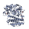
| Title | OvsA from Halomonas utahensis, an ovoselenol-biosynthetic selenoxide synthase in complex with histidine | |||||||||
 Components Components | Selenoxide synthase OvsA | |||||||||
 Keywords Keywords | OXIDOREDUCTASE / ovoselenol / selenium / selenoxide / nonheme iron | |||||||||
| Function / homology | : / FORMIC ACID / HISTIDINE Function and homology information Function and homology information | |||||||||
| Biological species |  Halomonas utahensis (bacteria) Halomonas utahensis (bacteria) | |||||||||
| Method |  X-RAY DIFFRACTION / SYNCHROTRON / MOLECULAR REPLACEMENT / Resolution: 2.72 Å X-RAY DIFFRACTION / SYNCHROTRON / MOLECULAR REPLACEMENT / Resolution: 2.72 Å | |||||||||
 Authors Authors | Ireland, K.A. / Davis, K.M. | |||||||||
| Funding support |  United States, 2items United States, 2items
| |||||||||
 Citation Citation | Journal: Nat.Chem. / Year: 2024 Title: Discovery of the selenium-containing antioxidant ovoselenol derived from convergent evolution. Authors: Kayrouz, C.M. / Ireland, K.A. / Ying, V.Y. / Davis, K.M. / Seyedsayamdost, M.R. | |||||||||
| History |
|
- Structure visualization
| Structure viewer | Molecule: MolmilJmol/JSmol |
|---|
- Downloads & links
-Download
| PDBx/mmCIF format | 8u41.cif.gz | 210.7 KB | Display | PDBx/mmCIF format |
|---|---|---|---|---|
| PDB format | pdb8u41.ent.gz | 152.6 KB | Display | PDB format |
| PDBx/mmJSON format | 8u41.json.gz | Tree view | PDBx/mmJSON format | |
| Others |  Other downloads Other downloads |
-Validation report
| Arichive directory | https://data.pdbj.org/pub/pdb/validation_reports/u4/8u41ftp://data.pdbj.org/pub/pdb/validation_reports/u4/8u41 | HTTPS FTP |
|---|
-Related structure data

-Links
 PDBj
PDBj
- Assembly
| Deposited unit | 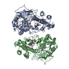
| ||||||||||||
|---|---|---|---|---|---|---|---|---|---|---|---|---|---|
| 1 | 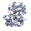
| ||||||||||||
| 2 | 
| ||||||||||||
| Unit cell |
|
-Components
-Protein , 1 types, 2 molecules AB
| #1: Protein | Mass: 54745.898 Da / Num. of mol.: 2 Source method: isolated from a genetically manipulated source Source: (gene. exp.) Halomonas utahensis (bacteria) / Strain: DSM 3051 / Gene: CK501_04640 / Production host: |
|---|
-Non-polymers , 5 types, 93 molecules 


| #2: Chemical |  Mass: 55.845 Da / Num. of mol.: 2 / Source method: obtained synthetically / Formula: Fe Mass: 55.845 Da / Num. of mol.: 2 / Source method: obtained synthetically / Formula: Fe#3: Chemical |  Mass: 22.990 Da / Num. of mol.: 2 / Source method: obtained synthetically / Formula: Na Mass: 22.990 Da / Num. of mol.: 2 / Source method: obtained synthetically / Formula: Na#4: Chemical |  Mass: 46.025 Da / Num. of mol.: 2 / Source method: obtained synthetically / Formula: CH2O2 Mass: 46.025 Da / Num. of mol.: 2 / Source method: obtained synthetically / Formula: CH2O2#5: Chemical |  Type: L-peptide linking / Mass: 156.162 Da / Num. of mol.: 2 / Source method: obtained synthetically / Formula: C6H10N3O2 / Feature type: SUBJECT OF INVESTIGATION Type: L-peptide linking / Mass: 156.162 Da / Num. of mol.: 2 / Source method: obtained synthetically / Formula: C6H10N3O2 / Feature type: SUBJECT OF INVESTIGATION#6: Water | ChemComp-HOH / | Mass: 18.015 Da / Num. of mol.: 85 / Source method: isolated from a natural source / Formula: H2O |
|---|
-Details
| Has ligand of interest | Y |
|---|---|
| Has protein modification | N |
-Experimental details
-Experiment
| Experiment | Method: X-RAY DIFFRACTION / Number of used crystals: 1 |
|---|
- Sample preparation
| Crystal | Density Matthews: 4.26 Å3/Da / Density % sol: 71.2 % / Description: Clear hexagonal rods |
|---|---|
| Crystal grow | Temperature: 293 K / Method: vapor diffusion, sitting drop / pH: 4.8 / Details: 0.1 M sodium acetate pH 4.8, 3.9 M sodium formate |
-Data collection
| Diffraction | Mean temperature: 100 K / Serial crystal experiment: N |
|---|---|
| Diffraction source | Source: SYNCHROTRON / Site: APS / Beamline: 21-ID-F / Wavelength: 0.9787 Å |
| Detector | Type: RAYONIX MX-300 / Detector: CCD / Date: Feb 18, 2023 |
| Radiation | Monochromator: C(111) / Protocol: SINGLE WAVELENGTH / Monochromatic (M) / Laue (L): M / Scattering type: x-ray |
| Radiation wavelength | Wavelength: 0.9787 Å / Relative weight: 1 |
| Reflection | Resolution: 2.72→69.34 Å / Num. obs: 47873 / % possible obs: 99.93 % / Redundancy: 2 % / Biso Wilson estimate: 74.79 Å2 / CC1/2: 0.999 / CC star: 1 / Rmerge(I) obs: 0.02925 / Rpim(I) all: 0.02925 / Rrim(I) all: 0.04136 / Net I/σ(I): 16.89 |
| Reflection shell | Resolution: 2.72→2.817 Å / Redundancy: 2 % / Rmerge(I) obs: 0.5307 / Mean I/σ(I) obs: 1.5 / Num. unique obs: 4740 / CC1/2: 0.654 / CC star: 0.889 / Rpim(I) all: 0.5307 / Rrim(I) all: 0.7505 / % possible all: 99.92 |
- Processing
| Software |
| |||||||||||||||||||||||||||||||||||||||||||||||||||||||||||||||||||||||||||||||||||||||||||||||||||||||||
|---|---|---|---|---|---|---|---|---|---|---|---|---|---|---|---|---|---|---|---|---|---|---|---|---|---|---|---|---|---|---|---|---|---|---|---|---|---|---|---|---|---|---|---|---|---|---|---|---|---|---|---|---|---|---|---|---|---|---|---|---|---|---|---|---|---|---|---|---|---|---|---|---|---|---|---|---|---|---|---|---|---|---|---|---|---|---|---|---|---|---|---|---|---|---|---|---|---|---|---|---|---|---|---|---|---|---|
| Refinement | Method to determine structure: MOLECULAR REPLACEMENT / Resolution: 2.72→69.34 Å / SU ML: 0.468 / Cross valid method: FREE R-VALUE / σ(F): 1.34 / Phase error: 31.0053 Stereochemistry target values: GeoStd + Monomer Library + CDL v1.2
| |||||||||||||||||||||||||||||||||||||||||||||||||||||||||||||||||||||||||||||||||||||||||||||||||||||||||
| Solvent computation | Shrinkage radii: 0.9 Å / VDW probe radii: 1.1 Å / Solvent model: FLAT BULK SOLVENT MODEL | |||||||||||||||||||||||||||||||||||||||||||||||||||||||||||||||||||||||||||||||||||||||||||||||||||||||||
| Displacement parameters | Biso mean: 78.6 Å2 | |||||||||||||||||||||||||||||||||||||||||||||||||||||||||||||||||||||||||||||||||||||||||||||||||||||||||
| Refinement step | Cycle: LAST / Resolution: 2.72→69.34 Å
| |||||||||||||||||||||||||||||||||||||||||||||||||||||||||||||||||||||||||||||||||||||||||||||||||||||||||
| Refine LS restraints |
| |||||||||||||||||||||||||||||||||||||||||||||||||||||||||||||||||||||||||||||||||||||||||||||||||||||||||
| LS refinement shell |
|
