 Movie
Movie Controller

[English] 日本語
 Yorodumi
- PDB-8u2q: Crystal Structure of Glycine--tRNA ligase active site chimera fro... -
+ Open data

- Basic information
| Entry | Database: PDB / ID: 8u2q | |||||||||
|---|---|---|---|---|---|---|---|---|---|---|
| Title | Crystal Structure of Glycine--tRNA ligase active site chimera from Mycobacterium thermoresistibile/tuberculosis (G5A bound) | |||||||||
 Components Components | Glycine--tRNA ligase | |||||||||
 Keywords Keywords | TRANSFERASE / SSGCID / STRUCTURAL GENOMICS / SEATTLE STRUCTURAL GENOMICS CENTER FOR INFECTIOUS DISEASE / Glycine--tRNA ligase | |||||||||
| Function / homology |  Function and homology information Function and homology informationglycyl-tRNA aminoacylation / glycine-tRNA ligase / microvesicle / glycine-tRNA ligase activity / bis(5'-nucleosyl)-tetraphosphatase (asymmetrical) activity / diadenosine tetraphosphate biosynthetic process / protein dimerization activity / extracellular exosome / ATP binding / metal ion binding / cytosol Similarity search - Function | |||||||||
| Biological species |  Mycolicibacterium thermoresistibile ATCC 19527 (bacteria) Mycolicibacterium thermoresistibile ATCC 19527 (bacteria) | |||||||||
| Method |  X-RAY DIFFRACTION / SYNCHROTRON / MOLECULAR REPLACEMENT / Resolution: 2.45 Å X-RAY DIFFRACTION / SYNCHROTRON / MOLECULAR REPLACEMENT / Resolution: 2.45 Å | |||||||||
 Authors Authors | Seattle Structural Genomics Center for Infectious Disease / Seattle Structural Genomics Center for Infectious Disease (SSGCID) | |||||||||
| Funding support |  United States, 2items United States, 2items
| |||||||||
 Citation Citation | Journal: To be published Title: Crystal Structure of Glycine--tRNA ligase active site chimera from Mycobacterium thermoresistibile/tuberculosis (G5A bound) Authors: Seibold, S. / Lovell, S. / Battaile, K.P. / DeRocher, A. | |||||||||
| History |
|
- Structure visualization
| Structure viewer | Molecule: MolmilJmol/JSmol |
|---|
- Downloads & links
-Download
| PDBx/mmCIF format | 8u2q.cif.gz | 352.1 KB | Display | PDBx/mmCIF format |
|---|---|---|---|---|
| PDB format | pdb8u2q.ent.gz | 285 KB | Display | PDB format |
| PDBx/mmJSON format | 8u2q.json.gz | Tree view | PDBx/mmJSON format | |
| Others |  Other downloads Other downloads |
-Validation report
| Arichive directory | https://data.pdbj.org/pub/pdb/validation_reports/u2/8u2qftp://data.pdbj.org/pub/pdb/validation_reports/u2/8u2q | HTTPS FTP |
|---|
-Related structure data
| Similar structure data |
|---|
-Links
 PDBj
PDBj
- Assembly
| Deposited unit | 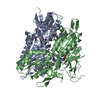
| ||||||||
|---|---|---|---|---|---|---|---|---|---|
| 1 |
| ||||||||
| Unit cell |
|
-Components
-Protein , 1 types, 2 molecules AB
| #1: Protein | Mass: 54663.113 Da / Num. of mol.: 2 / Fragment: V122, P123 deletion / Mutation: H97Y, Q99A, D121G Source method: isolated from a genetically manipulated source Source: (gene. exp.) Mycolicibacterium thermoresistibile ATCC 19527 (bacteria)Gene: glyQS / Plasmid: MythA.19107.a.UX11 / Production host: |
|---|
-Non-polymers , 6 types, 115 molecules 


| #2: Chemical |  Mass: 22.990 Da / Num. of mol.: 2 / Source method: obtained synthetically / Formula: Na Mass: 22.990 Da / Num. of mol.: 2 / Source method: obtained synthetically / Formula: Na#3: Chemical |  Mass: 35.453 Da / Num. of mol.: 2 / Source method: obtained synthetically / Formula: Cl Mass: 35.453 Da / Num. of mol.: 2 / Source method: obtained synthetically / Formula: Cl#4: Chemical | ChemComp-EDO / |  Mass: 62.068 Da / Num. of mol.: 1 / Source method: obtained synthetically / Formula: C2H6O2 Mass: 62.068 Da / Num. of mol.: 1 / Source method: obtained synthetically / Formula: C2H6O2#5: Chemical |  Mass: 403.371 Da / Num. of mol.: 2 / Source method: obtained synthetically / Formula: C12H17N7O7S / Feature type: SUBJECT OF INVESTIGATION Mass: 403.371 Da / Num. of mol.: 2 / Source method: obtained synthetically / Formula: C12H17N7O7S / Feature type: SUBJECT OF INVESTIGATION#6: Chemical | ChemComp-ZN / |  Mass: 65.409 Da / Num. of mol.: 1 / Source method: obtained synthetically / Formula: Zn Mass: 65.409 Da / Num. of mol.: 1 / Source method: obtained synthetically / Formula: Zn#7: Water | ChemComp-HOH / | Mass: 18.015 Da / Num. of mol.: 107 / Source method: isolated from a natural source / Formula: H2O |
|---|
-Details
| Has ligand of interest | Y |
|---|
-Experimental details
-Experiment
| Experiment | Method: X-RAY DIFFRACTION / Number of used crystals: 1 |
|---|
- Sample preparation
| Crystal | Density Matthews: 3.29 Å3/Da / Density % sol: 62.63 % |
|---|---|
| Crystal grow | Temperature: 291 K / Method: vapor diffusion, sitting drop / pH: 7.5 Details: Index F12: 25% (w/v) Polyethylene glycol 3,350, 0.1 M HEPES pH 7.5, 0.2 M Sodium chloride. MythA.19107.a.UX11.PW39208 at 20.4 mg/mL. 2mM G5A added prior to crystallization. Plate 13386 F12 ...Details: Index F12: 25% (w/v) Polyethylene glycol 3,350, 0.1 M HEPES pH 7.5, 0.2 M Sodium chloride. MythA.19107.a.UX11.PW39208 at 20.4 mg/mL. 2mM G5A added prior to crystallization. Plate 13386 F12 drop 3. Puck: PSL-1203, Cryo: 20% Glycerol + 80% Crystallant. |
-Data collection
| Diffraction | Mean temperature: 100 K / Serial crystal experiment: N |
|---|---|
| Diffraction source | Source: SYNCHROTRON / Site: NSLS-II / Beamline: 19-ID / Wavelength: 0.9785 Å |
| Detector | Type: DECTRIS EIGER2 XE 9M / Detector: PIXEL / Date: Aug 6, 2023 |
| Radiation | Monochromator: Double Crystal Si 111 / Protocol: SINGLE WAVELENGTH / Monochromatic (M) / Laue (L): M / Scattering type: x-ray |
| Radiation wavelength | Wavelength: 0.9785 Å / Relative weight: 1 |
| Reflection | Resolution: 2.45→48.08 Å / Num. obs: 52055 / % possible obs: 99.6 % / Redundancy: 3.5 % / CC1/2: 0.997 / Rmerge(I) obs: 0.086 / Rpim(I) all: 0.053 / Rrim(I) all: 0.101 / Χ2: 0.94 / Net I/σ(I): 9.8 / Num. measured all: 182866 |
| Reflection shell | Resolution: 2.45→2.53 Å / % possible obs: 99.8 % / Redundancy: 3.7 % / Rmerge(I) obs: 0.887 / Num. measured all: 16592 / Num. unique obs: 4494 / CC1/2: 0.756 / Rpim(I) all: 0.533 / Rrim(I) all: 1.037 / Χ2: 0.98 / Net I/σ(I) obs: 1.9 |
- Processing
| Software |
| |||||||||||||||||||||||||||||||||||||||||||||||||||||||||||||||||||||||||||||||||||||||||||||||||||||||||||||||||||||||||||||||||||||||||||||||||||||||||||||||||||||||||||||||||||||||||||||||||||||||||||||||||||||||||||||||||
|---|---|---|---|---|---|---|---|---|---|---|---|---|---|---|---|---|---|---|---|---|---|---|---|---|---|---|---|---|---|---|---|---|---|---|---|---|---|---|---|---|---|---|---|---|---|---|---|---|---|---|---|---|---|---|---|---|---|---|---|---|---|---|---|---|---|---|---|---|---|---|---|---|---|---|---|---|---|---|---|---|---|---|---|---|---|---|---|---|---|---|---|---|---|---|---|---|---|---|---|---|---|---|---|---|---|---|---|---|---|---|---|---|---|---|---|---|---|---|---|---|---|---|---|---|---|---|---|---|---|---|---|---|---|---|---|---|---|---|---|---|---|---|---|---|---|---|---|---|---|---|---|---|---|---|---|---|---|---|---|---|---|---|---|---|---|---|---|---|---|---|---|---|---|---|---|---|---|---|---|---|---|---|---|---|---|---|---|---|---|---|---|---|---|---|---|---|---|---|---|---|---|---|---|---|---|---|---|---|---|---|---|---|---|---|---|---|---|---|---|---|---|---|---|---|---|---|
| Refinement | Method to determine structure: MOLECULAR REPLACEMENT / Resolution: 2.45→41.53 Å / SU ML: 0.3 / Cross valid method: FREE R-VALUE / σ(F): 1.34 / Phase error: 23.49 / Stereochemistry target values: ML
| |||||||||||||||||||||||||||||||||||||||||||||||||||||||||||||||||||||||||||||||||||||||||||||||||||||||||||||||||||||||||||||||||||||||||||||||||||||||||||||||||||||||||||||||||||||||||||||||||||||||||||||||||||||||||||||||||
| Solvent computation | Shrinkage radii: 0.9 Å / VDW probe radii: 1.1 Å / Solvent model: FLAT BULK SOLVENT MODEL | |||||||||||||||||||||||||||||||||||||||||||||||||||||||||||||||||||||||||||||||||||||||||||||||||||||||||||||||||||||||||||||||||||||||||||||||||||||||||||||||||||||||||||||||||||||||||||||||||||||||||||||||||||||||||||||||||
| Refinement step | Cycle: LAST / Resolution: 2.45→41.53 Å
| |||||||||||||||||||||||||||||||||||||||||||||||||||||||||||||||||||||||||||||||||||||||||||||||||||||||||||||||||||||||||||||||||||||||||||||||||||||||||||||||||||||||||||||||||||||||||||||||||||||||||||||||||||||||||||||||||
| Refine LS restraints |
| |||||||||||||||||||||||||||||||||||||||||||||||||||||||||||||||||||||||||||||||||||||||||||||||||||||||||||||||||||||||||||||||||||||||||||||||||||||||||||||||||||||||||||||||||||||||||||||||||||||||||||||||||||||||||||||||||
| LS refinement shell |
| |||||||||||||||||||||||||||||||||||||||||||||||||||||||||||||||||||||||||||||||||||||||||||||||||||||||||||||||||||||||||||||||||||||||||||||||||||||||||||||||||||||||||||||||||||||||||||||||||||||||||||||||||||||||||||||||||
| Refinement TLS params. | Method: refined / Refine-ID: X-RAY DIFFRACTION
| |||||||||||||||||||||||||||||||||||||||||||||||||||||||||||||||||||||||||||||||||||||||||||||||||||||||||||||||||||||||||||||||||||||||||||||||||||||||||||||||||||||||||||||||||||||||||||||||||||||||||||||||||||||||||||||||||
| Refinement TLS group |
|
