 Movie
Movie Controller

[English] 日本語
 Yorodumi
- PDB-8u1q: A mechanistic understanding of protective influenza B neuraminida... -
+ Open data

- Basic information
| Entry | Database: PDB / ID: 8u1q | ||||||
|---|---|---|---|---|---|---|---|
| Title | A mechanistic understanding of protective influenza B neuraminidase mAbs at the airway interface | ||||||
 Components Components |
| ||||||
 Keywords Keywords | HYDROLASE / Neuraminidase Sialidase | ||||||
| Function / homology |  Function and homology information Function and homology informationexo-alpha-sialidase / exo-alpha-sialidase activity / carbohydrate metabolic process / host cell plasma membrane / virion membrane / membrane / metal ion binding Similarity search - Function | ||||||
| Biological species |  Influenza B virus Influenza B virus Homo sapiens (human) Homo sapiens (human) | ||||||
| Method | ELECTRON MICROSCOPY / single particle reconstruction / cryo EM / Resolution: 3.36 Å | ||||||
 Authors Authors | Ferguson, J.A. / Oeverdieck, S. / Ward, A.B. | ||||||
| Funding support |  United States, 1items United States, 1items
| ||||||
 Citation Citation | Journal: Immunity / Year: 2024 Title: Isolation of human antibodies against influenza B neuraminidase and mechanisms of protection at the airway interface. Authors: Rachael M Wolters / James A Ferguson / Ivette A Nuñez / Elaine E Chen / Ty Sornberger / Luke Myers / Svearike Oeverdieck / Sai Sundar Rajan Raghavan / Chandrahaas Kona / Laura S Handal / ...Authors: Rachael M Wolters / James A Ferguson / Ivette A Nuñez / Elaine E Chen / Ty Sornberger / Luke Myers / Svearike Oeverdieck / Sai Sundar Rajan Raghavan / Chandrahaas Kona / Laura S Handal / Trevor E Esilu / Edgar Davidson / Benjamin J Doranz / Taylor B Engdahl / Nurgun Kose / Lauren E Williamson / C Buddy Creech / Katherine N Gibson-Corley / Andrew B Ward / James E Crowe / Abstract: Influenza B viruses (IBVs) comprise a substantial portion of the circulating seasonal human influenza viruses. Here, we describe the isolation of human monoclonal antibodies (mAbs) that recognized ...Influenza B viruses (IBVs) comprise a substantial portion of the circulating seasonal human influenza viruses. Here, we describe the isolation of human monoclonal antibodies (mAbs) that recognized the IBV neuraminidase (NA) glycoprotein from an individual following seasonal vaccination. Competition-binding experiments suggested the antibodies recognized two major antigenic sites. One group, which included mAb FluB-393, broadly inhibited IBV NA sialidase activity, protected prophylactically in vivo, and bound to the lateral corner of NA. The second group contained an active site mAb, FluB-400, that broadly inhibited IBV NA sialidase activity and virus replication in vitro in primary human respiratory epithelial cell cultures and protected against IBV in vivo when administered systemically or intranasally. Overall, the findings described here shape our mechanistic understanding of the human immune response to the IBV NA glycoprotein through the demonstration of two mAb delivery routes for protection against IBV and the identification of potential IBV therapeutic candidates. | ||||||
| History |
|
- Structure visualization
| Structure viewer | Molecule: MolmilJmol/JSmol |
|---|
- Downloads & links
-Download
| PDBx/mmCIF format | 8u1q.cif.gz | 135.7 KB | Display | PDBx/mmCIF format |
|---|---|---|---|---|
| PDB format | pdb8u1q.ent.gz | 100.4 KB | Display | PDB format |
| PDBx/mmJSON format | 8u1q.json.gz | Tree view | PDBx/mmJSON format | |
| Others |  Other downloads Other downloads |
-Validation report
| Arichive directory | https://data.pdbj.org/pub/pdb/validation_reports/u1/8u1qftp://data.pdbj.org/pub/pdb/validation_reports/u1/8u1q | HTTPS FTP |
|---|
-Related structure data
| Related structure data |  41824MC 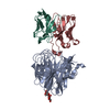 8u1cC 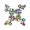 8u1sC M: map data used to model this data C: citing same article ( |
|---|---|
| Similar structure data |


-Links
 PDBj
PDBj


- Assembly
| Deposited unit | 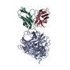
|
|---|---|
| 1 | 
|
| 2 |
|
| 3 | 
|
| Symmetry | Point symmetry: (Schoenflies symbol: C4 (4 fold cyclic)) |
-Components
| #1: Protein | Mass: 51104.258 Da / Num. of mol.: 1 Source method: isolated from a genetically manipulated source Source: (gene. exp.) Influenza B virus (B/Iowa/06/2017) / Gene: NA / Production host:  |
|---|---|
| #2: Antibody | Mass: 13139.580 Da / Num. of mol.: 1 Source method: isolated from a genetically manipulated source Source: (gene. exp.) Homo sapiens (human) / Cell line (production host): ExpiCHO / Production host:   Cricetulus griseus (Chinese hamster) Cricetulus griseus (Chinese hamster) |
| #3: Antibody | Mass: 11627.834 Da / Num. of mol.: 1 Source method: isolated from a genetically manipulated source Source: (gene. exp.) Homo sapiens (human) / Cell line (production host): ExpiCHO / Production host: Cricetulus griseus (Chinese hamster) |
| #4: Polysaccharide | 2-acetamido-2-deoxy-beta-D-glucopyranose-(1-4)-2-acetamido-2-deoxy-beta-D-glucopyranose Source method: isolated from a genetically manipulated source |
| Has ligand of interest | Y |
| Has protein modification | Y |
-Experimental details
-Experiment
| Experiment | Method: ELECTRON MICROSCOPY |
|---|---|
| EM experiment | Aggregation state: PARTICLE / 3D reconstruction method: single particle reconstruction |
- Sample preparation
| Component | Name: Complex of human mAb-fv domain bound to influenza B neuraminidase, from a public database. Type: COMPLEX / Entity ID: #1-#3 / Source: MULTIPLE SOURCES | ||||||||||||||||||
|---|---|---|---|---|---|---|---|---|---|---|---|---|---|---|---|---|---|---|---|
| Molecular weight | Units: MEGADALTONS / Experimental value: NO | ||||||||||||||||||
| Source (natural) | Organism: Homo sapiens (human) | ||||||||||||||||||
| Source (recombinant) |
| ||||||||||||||||||
| Buffer solution | pH: 7.4 / Details: TBS | ||||||||||||||||||
| Specimen | Conc.: 0.4 mg/ml / Embedding applied: NO / Shadowing applied: NO / Staining applied: NO / Vitrification applied: YES | ||||||||||||||||||
| Vitrification | Instrument: FEI VITROBOT MARK III / Cryogen name: ETHANE / Humidity: 100 % / Chamber temperature: 298 K |
- Electron microscopy imaging
| Experimental equipment |  Model: Talos Arctica / Image courtesy: FEI Company |
|---|---|
| Microscopy | Model: FEI TALOS ARCTICA |
| Electron gun | Electron source:  FIELD EMISSION GUN / Accelerating voltage: 200 kV / Illumination mode: FLOOD BEAM FIELD EMISSION GUN / Accelerating voltage: 200 kV / Illumination mode: FLOOD BEAM |
| Electron lens | Mode: BRIGHT FIELD / Nominal magnification: 36000 X / Calibrated magnification: 36000 X / Nominal defocus max: 700 nm / Nominal defocus min: 700 nm / Calibrated defocus min: 2000 nm / Calibrated defocus max: 2000 nm / Cs: 2.7 mm / C2 aperture diameter: 100 µm |
| Specimen holder | Cryogen: NITROGEN / Specimen holder model: OTHER |
| Image recording | Electron dose: 50.43 e/Å2 / Detector mode: COUNTING / Film or detector model: GATAN K2 SUMMIT (4k x 4k) / Num. of grids imaged: 1 / Num. of real images: 2559 |
| Image scans | Width: 4048 / Height: 4048 |
- Processing
| EM software |
| ||||||||||||||||||||||||||||||||||||||||
|---|---|---|---|---|---|---|---|---|---|---|---|---|---|---|---|---|---|---|---|---|---|---|---|---|---|---|---|---|---|---|---|---|---|---|---|---|---|---|---|---|---|
| CTF correction | Type: NONE | ||||||||||||||||||||||||||||||||||||||||
| 3D reconstruction | Resolution: 3.36 Å / Resolution method: FSC 0.143 CUT-OFF / Num. of particles: 335369 / Symmetry type: POINT | ||||||||||||||||||||||||||||||||||||||||
| Atomic model building |
| ||||||||||||||||||||||||||||||||||||||||
| Refinement | Cross valid method: NONE Stereochemistry target values: GeoStd + Monomer Library + CDL v1.2 | ||||||||||||||||||||||||||||||||||||||||
| Displacement parameters | Biso mean: 45.01 Å2 | ||||||||||||||||||||||||||||||||||||||||
| Refine LS restraints |
|
