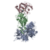
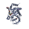
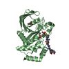
Journal: Commun Chem / Year: 2024 Title: Mechanistic insights into a heterobifunctional degrader-induced PTPN2/N1 complex. Authors: Qi Hao / Manoj K Rathinaswamy / Kelly L Klinge / Matthew Bratkowski / Amirhossein Mafi / Christina K Baumgartner / Keith M Hamel / Gesine K Veits / Rinku Jain / Claudio Catalano / Mark ...Authors: Qi Hao / Manoj K Rathinaswamy / Kelly L Klinge / Matthew Bratkowski / Amirhossein Mafi / Christina K Baumgartner / Keith M Hamel / Gesine K Veits / Rinku Jain / Claudio Catalano / Mark Fitzgerald / Alexander W Hird / Eunice Park / Harit U Vora / James A Henderson / Kenton Longenecker / Charles W Hutchins / Wei Qiu / Giovanna Scapin / Qi Sun / Vincent S Stoll / Chaohong Sun / Ping Li / Dan Eaton / David Stokoe / Stewart L Fisher / Christopher G Nasveschuk / Marcia Paddock / Michael E Kort / Abstract: PTPN2 (protein tyrosine phosphatase non-receptor type 2, or TC-PTP) and PTPN1 are attractive immuno-oncology targets, with the deletion of Ptpn1 and Ptpn2 improving response to immunotherapy in ...PTPN2 (protein tyrosine phosphatase non-receptor type 2, or TC-PTP) and PTPN1 are attractive immuno-oncology targets, with the deletion of Ptpn1 and Ptpn2 improving response to immunotherapy in disease models. Targeted protein degradation has emerged as a promising approach to drug challenging targets including phosphatases. We developed potent PTPN2/N1 dual heterobifunctional degraders (Cmpd-1 and Cmpd-2) which facilitate efficient complex assembly with E3 ubiquitin ligase CRL4, and mediate potent PTPN2/N1 degradation in cells and mice. To provide mechanistic insights into the cooperative complex formation introduced by degraders, we employed a combination of structural approaches. Our crystal structure reveals how PTPN2 is recognized by the tri-substituted thiophene moiety of the degrader. We further determined a high-resolution structure of DDB1-CRBN/Cmpd-1/PTPN2 using single-particle cryo-electron microscopy (cryo-EM). This structure reveals that the degrader induces proximity between CRBN and PTPN2, albeit the large conformational heterogeneity of this ternary complex. The molecular dynamic (MD)-simulations constructed based on the cryo-EM structure exhibited a large rigid body movement of PTPN2 and illustrated the dynamic interactions between PTPN2 and CRBN. Together, our study demonstrates the development of PTPN2/N1 heterobifunctional degraders with potential applications in cancer immunotherapy. Furthermore, the developed structural workflow could help to understand the dynamic nature of degrader-induced cooperative ternary complexes.
In the structure databanks used in Yorodumi, some data are registered as the other names, "COVID-19 virus" and "2019-nCoV". Here are the details of the virus and the list of structure data.
Jan 31, 2019. EMDB accession codes are about to change! (news from PDBe EMDB page)
EMDB accession codes are about to change! (news from PDBe EMDB page)
The allocation of 4 digits for EMDB accession codes will soon come to an end. Whilst these codes will remain in use, new EMDB accession codes will include an additional digit and will expand incrementally as the available range of codes is exhausted. The current 4-digit format prefixed with “EMD-” (i.e. EMD-XXXX) will advance to a 5-digit format (i.e. EMD-XXXXX), and so on. It is currently estimated that the 4-digit codes will be depleted around Spring 2019, at which point the 5-digit format will come into force.
The EM Navigator/Yorodumi systems omit the EMD- prefix.
Related info.:Q: What is EMD? / ID/Accession-code notation in Yorodumi/EM Navigator
Yorodumi is a browser for structure data from EMDB, PDB, SASBDB, etc.
This page is also the successor to EM Navigator detail page, and also detail information page/front-end page for Omokage search.
The word "yorodu" (or yorozu) is an old Japanese word meaning "ten thousand". "mi" (miru) is to see.
Related info.:EMDB / PDB / SASBDB / Comparison of 3 databanks / Yorodumi Search / Aug 31, 2016. New EM Navigator & Yorodumi / Yorodumi Papers / Jmol/JSmol / Function and homology information / Changes in new EM Navigator and Yorodumi
 Movie
Movie Controller


 Open data
 Basic information
 Components
 Keywords
 Function and homology information
 Homo sapiens (human)
 X-RAY DIFFRACTION /
  Authors
 Citation
 Structure visualization
 Downloads & links
 Other downloads
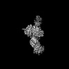
 PDBj
PDBj


 Assembly
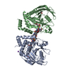

 Spodoptera frugiperda (fall armyworm) / References: UniProt: P17706, protein-tyrosine-phosphatase
 Mass: 59.044 Da / Num. of mol.: 2 / Source method: obtained synthetically / Formula: C2H3O2
 Mass: 22.990 Da / Num. of mol.: 1 / Source method: obtained synthetically / Formula: Na
 Mass: 18.015 Da / Num. of mol.: 385 / Source method: isolated from a natural source / Formula: H2O
 Sample preparation
 Processing