 Movie
Movie Controller


+ Open data

- Basic information
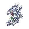
| Entry | Database: PDB / ID: 8two | ||||||
|---|---|---|---|---|---|---|---|
| Title | AvrB bound with UDP and RIN4_T166-Rha | ||||||
 Components Components |
| ||||||
 Keywords Keywords | TRANSFERASE / Protein complex | ||||||
| Function / homology | Avirulence B/C / Avirulence B/C superfamily / : / Avirulence protein / extracellular region / alpha-L-rhamnopyranose / URIDINE-5'-DIPHOSPHATE / Avirulence protein B Function and homology information Function and homology information | ||||||
| Biological species |  Pseudomonas syringae (bacteria) Pseudomonas syringae (bacteria) | ||||||
| Method |  X-RAY DIFFRACTION / SYNCHROTRON / MOLECULAR REPLACEMENT / Resolution: 2.09 Å X-RAY DIFFRACTION / SYNCHROTRON / MOLECULAR REPLACEMENT / Resolution: 2.09 Å | ||||||
 Authors Authors | Peng, W. / Orth, K. | ||||||
| Funding support |  United States, 1items United States, 1items
| ||||||
 Citation Citation | Journal: Sci Adv / Year: 2024 Title: Pseudomonas effector AvrB is a glycosyltransferase that rhamnosylates plant guardee protein RIN4. Authors: Peng, W. / Garcia, N. / Servage, K.A. / Kohler, J.J. / Ready, J.M. / Tomchick, D.R. / Fernandez, J. / Orth, K. | ||||||
| History |
|
- Structure visualization
| Structure viewer | Molecule: MolmilJmol/JSmol |
|---|
- Downloads & links
-Download
| PDBx/mmCIF format | 8two.cif.gz | 197.4 KB | Display | PDBx/mmCIF format |
|---|---|---|---|---|
| PDB format | pdb8two.ent.gz | 158 KB | Display | PDB format |
| PDBx/mmJSON format | 8two.json.gz | Tree view | PDBx/mmJSON format | |
| Others |  Other downloads Other downloads |
-Validation report
| Arichive directory | https://data.pdbj.org/pub/pdb/validation_reports/tw/8twoftp://data.pdbj.org/pub/pdb/validation_reports/tw/8two | HTTPS FTP |
|---|
-Related structure data


-Links
 PDBj
PDBj


- Assembly
| Deposited unit | 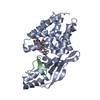
| ||||||||
|---|---|---|---|---|---|---|---|---|---|
| 1 |
| ||||||||
| Unit cell |
|
-Components
| #1: Protein | Mass: 36262.359 Da / Num. of mol.: 1 Source method: isolated from a genetically manipulated source Source: (gene. exp.) Pseudomonas syringae (bacteria) / Gene: avrB / Production host: |
|---|---|
| #2: Protein/peptide | Mass: 3428.589 Da / Num. of mol.: 1 Source method: isolated from a genetically manipulated source Source: (gene. exp.) |
| #3: Chemical | ChemComp-UDP /   Type: RNA linking / Mass: 404.161 Da / Num. of mol.: 1 / Source method: obtained synthetically / Formula: C9H14N2O12P2 / Feature type: SUBJECT OF INVESTIGATION / Comment: UDP*YM Type: RNA linking / Mass: 404.161 Da / Num. of mol.: 1 / Source method: obtained synthetically / Formula: C9H14N2O12P2 / Feature type: SUBJECT OF INVESTIGATION / Comment: UDP*YM |
| #4: Sugar | ChemComp-RAM /   Type: L-saccharide, alpha linking / Mass: 164.156 Da / Num. of mol.: 1 / Source method: obtained synthetically / Formula: C6H12O5 / Feature type: SUBJECT OF INVESTIGATION Type: L-saccharide, alpha linking / Mass: 164.156 Da / Num. of mol.: 1 / Source method: obtained synthetically / Formula: C6H12O5 / Feature type: SUBJECT OF INVESTIGATION |
| Has ligand of interest | Y |
-Experimental details
-Experiment
| Experiment | Method: X-RAY DIFFRACTION / Number of used crystals: 1 |
|---|
- Sample preparation
| Crystal grow | Temperature: 293 K / Method: vapor diffusion, hanging drop / Details: 100 mM Tris (pH 7.5~7.8), 27%~32% PEG 550 MME |
|---|
-Data collection
| Diffraction | Mean temperature: 100 K / Serial crystal experiment: N |
|---|---|
| Diffraction source | Source: SYNCHROTRON / Site: ALS / Beamline: 2.0.1 / Wavelength: 1.0358 Å |
| Detector | Type: DECTRIS PILATUS 2M / Detector: PIXEL / Date: Jun 9, 2023 |
| Radiation | Protocol: SINGLE WAVELENGTH / Monochromatic (M) / Laue (L): M / Scattering type: x-ray |
| Radiation wavelength | Wavelength: 1.0358 Å / Relative weight: 1 |
| Reflection | Resolution: 2.09→52.15 Å / Num. obs: 16986 / % possible obs: 99.9 % / Redundancy: 6.3 % / Biso Wilson estimate: 49.65 Å2 / CC1/2: 0.997 / Net I/σ(I): 6.61 |
| Reflection shell | Resolution: 2.09→2.13 Å / Num. unique obs: 828 / CC1/2: 0.233 |
- Processing
| Software |
| |||||||||||||||||||||||||||||||||||||||||||||||||||||||||||||||||||||||||||||||||||||||||||
|---|---|---|---|---|---|---|---|---|---|---|---|---|---|---|---|---|---|---|---|---|---|---|---|---|---|---|---|---|---|---|---|---|---|---|---|---|---|---|---|---|---|---|---|---|---|---|---|---|---|---|---|---|---|---|---|---|---|---|---|---|---|---|---|---|---|---|---|---|---|---|---|---|---|---|---|---|---|---|---|---|---|---|---|---|---|---|---|---|---|---|---|---|
| Refinement | Method to determine structure: MOLECULAR REPLACEMENT / Resolution: 2.09→52.15 Å / SU ML: 0.5 / Cross valid method: FREE R-VALUE / σ(F): 1.33 / Phase error: 35.63 / Stereochemistry target values: ML
| |||||||||||||||||||||||||||||||||||||||||||||||||||||||||||||||||||||||||||||||||||||||||||
| Solvent computation | Shrinkage radii: 0.9 Å / VDW probe radii: 1.1 Å / Solvent model: FLAT BULK SOLVENT MODEL | |||||||||||||||||||||||||||||||||||||||||||||||||||||||||||||||||||||||||||||||||||||||||||
| Refinement step | Cycle: LAST / Resolution: 2.09→52.15 Å
| |||||||||||||||||||||||||||||||||||||||||||||||||||||||||||||||||||||||||||||||||||||||||||
| Refine LS restraints |
| |||||||||||||||||||||||||||||||||||||||||||||||||||||||||||||||||||||||||||||||||||||||||||
| LS refinement shell |
| |||||||||||||||||||||||||||||||||||||||||||||||||||||||||||||||||||||||||||||||||||||||||||
| Refinement TLS params. | Method: refined / Origin x: 19.4834 Å / Origin y: 22.8961 Å / Origin z: 9.8324 Å
| |||||||||||||||||||||||||||||||||||||||||||||||||||||||||||||||||||||||||||||||||||||||||||
| Refinement TLS group | Selection details: all |
